 Movie
Movie Controller

[English] 日本語
 Yorodumi
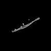
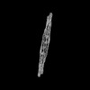
- PDB-8afl: Cryo-EM structure of crescentin filaments (wildtype, C1 symmetry ... -
+ Open data

- Basic information
| Entry | Database: PDB / ID: 8afl | ||||||
|---|---|---|---|---|---|---|---|
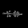
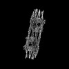
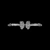
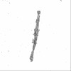
| Title | Cryo-EM structure of crescentin filaments (wildtype, C1 symmetry and small box) | ||||||
 Components Components |
| ||||||
 Keywords Keywords | STRUCTURAL PROTEIN / cytoskeleton / cell shape / intermediate filaments / coiled coil / assembly | ||||||
| Function / homology | :  Function and homology information Function and homology information | ||||||
| Biological species |  Caulobacter vibrioides (bacteria) Caulobacter vibrioides (bacteria) Camelidae (mammal) Camelidae (mammal) | ||||||
| Method | ELECTRON MICROSCOPY / single particle reconstruction / cryo EM / Resolution: 4.4 Å | ||||||
 Authors Authors | Liu, Y. / Lowe, J. | ||||||
| Funding support |  United Kingdom, 1items United Kingdom, 1items
| ||||||
 Citation Citation | Journal: Proc Natl Acad Sci U S A / Year: 2024 Title: Filament structure and subcellular organization of the bacterial intermediate filament-like protein crescentin. Authors: Yue Liu / Fusinita van den Ent / Jan Löwe / Abstract: The protein crescentin is required for the crescent shape of the freshwater bacterium (). Crescentin forms a filamentous structure on the inner, concave side of the curved cells. It shares features ...The protein crescentin is required for the crescent shape of the freshwater bacterium (). Crescentin forms a filamentous structure on the inner, concave side of the curved cells. It shares features with eukaryotic intermediate filament (IF) proteins, including the formation of static filaments based on long and parallel coiled coils, the protein's length, structural roles in cell and organelle shape determination and the presence of a coiled coil discontinuity called the "stutter." Here, we have used electron cryomicroscopy (cryo-EM) to determine the structure of the full-length protein and its filament, exploiting a crescentin-specific nanobody. The filament is formed by two strands, related by twofold symmetry, that each consist of two dimers, resulting in an octameric assembly. Crescentin subunits form longitudinal contacts head-to-head and tail-to-tail, making the entire filament non-polar. Using in vivo site-directed cysteine cross-linking, we demonstrated that contacts observed in the in vitro filament structure exist in cells. Electron cryotomography (cryo-ET) of cells expressing crescentin showed filaments on the concave side of the curved cells, close to the inner membrane, where they form a band. When comparing with current models of IF proteins and their filaments, which are also built from parallel coiled coil dimers and lack overall polarity, it emerges that IF proteins form head-to-tail longitudinal contacts in contrast to crescentin and hence several inter-dimer contacts in IFs have no equivalents in crescentin filaments. Our work supports the idea that intermediate filament-like proteins achieve their shared polymerization and mechanical properties through a variety of filament architectures. | ||||||
| History |
|
- Structure visualization
| Structure viewer | Molecule: MolmilJmol/JSmol |
|---|
- Downloads & links
-Download
| PDBx/mmCIF format | 8afl.cif.gz | 149.4 KB | Display | PDBx/mmCIF format |
|---|---|---|---|---|
| PDB format | pdb8afl.ent.gz | 86.4 KB | Display | PDB format |
| PDBx/mmJSON format | 8afl.json.gz | Tree view | PDBx/mmJSON format | |
| Others |  Other downloads Other downloads |
-Validation report
| Arichive directory | https://data.pdbj.org/pub/pdb/validation_reports/af/8aflftp://data.pdbj.org/pub/pdb/validation_reports/af/8afl | HTTPS FTP |
|---|
-Related structure data
| Related structure data |  15401MC  8afeC 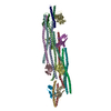 8afhC  8afmC  8ahlC 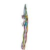 8aiaC 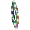 8aixC 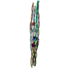 8ajbC M: map data used to model this data C: citing same article ( |
|---|---|
| Similar structure data |


-Links
 PDBj
PDBj

- Assembly
| Deposited unit | 
|
|---|---|
| 1 |
|
-Components
| #1: Protein | Mass: 49918.559 Da / Num. of mol.: 4 Source method: isolated from a genetically manipulated source Source: (gene. exp.) Caulobacter vibrioides (bacteria) / Gene: creS, KZH45_19550 / Production host: #2: Antibody | Mass: 101790.180 Da / Num. of mol.: 2 Source method: isolated from a genetically manipulated source Source: (gene. exp.) Camelidae (mammal) / Production host: Has protein modification | Y | |
|---|
-Experimental details
-Experiment
| Experiment | Method: ELECTRON MICROSCOPY |
|---|---|
| EM experiment | Aggregation state: FILAMENT / 3D reconstruction method: single particle reconstruction |
- Sample preparation
| Component | Name: Crescentin filaments in complex with megabodies / Type: COMPLEX / Entity ID: all / Source: RECOMBINANT |
|---|---|
| Molecular weight | Experimental value: NO |
| Source (natural) | Organism: Caulobacter vibrioides (bacteria) |
| Source (recombinant) | Organism: |
| Buffer solution | pH: 6.5 / Details: PIPES 25mM, 0.05% CHAPS, pH 6.5 |
| Specimen | Conc.: 2 mg/ml / Embedding applied: NO / Shadowing applied: NO / Staining applied: NO / Vitrification applied: YES |
| Specimen support | Grid material: GOLD / Grid mesh size: 300 divisions/in. / Grid type: UltrAuFoil R2/2 |
| Vitrification | Instrument: FEI VITROBOT MARK IV / Cryogen name: ETHANE / Humidity: 100 % / Chamber temperature: 283 K |
- Electron microscopy imaging
| Experimental equipment |  Model: Titan Krios / Image courtesy: FEI Company |
|---|---|
| Microscopy | Model: FEI TITAN KRIOS |
| Electron gun | Electron source:  FIELD EMISSION GUN / Accelerating voltage: 300 kV / Illumination mode: FLOOD BEAM FIELD EMISSION GUN / Accelerating voltage: 300 kV / Illumination mode: FLOOD BEAM |
| Electron lens | Mode: BRIGHT FIELD / Nominal magnification: 75000 X / Nominal defocus max: 3500 nm / Nominal defocus min: 800 nm / Cs: 2.7 mm / C2 aperture diameter: 50 µm / Alignment procedure: COMA FREE |
| Specimen holder | Cryogen: NITROGEN / Specimen holder model: FEI TITAN KRIOS AUTOGRID HOLDER / Temperature (max): 80 K / Temperature (min): 80 K |
| Image recording | Average exposure time: 10 sec. / Electron dose: 34 e/Å2 / Film or detector model: FEI FALCON IV (4k x 4k) / Num. of grids imaged: 2 |
- Processing
| Software |
| ||||||||||||||||||||||||||||||||||||
|---|---|---|---|---|---|---|---|---|---|---|---|---|---|---|---|---|---|---|---|---|---|---|---|---|---|---|---|---|---|---|---|---|---|---|---|---|---|
| EM software |
| ||||||||||||||||||||||||||||||||||||
| CTF correction | Type: PHASE FLIPPING AND AMPLITUDE CORRECTION | ||||||||||||||||||||||||||||||||||||
| Symmetry | Point symmetry: C1 (asymmetric) | ||||||||||||||||||||||||||||||||||||
| 3D reconstruction | Resolution: 4.4 Å / Resolution method: FSC 0.143 CUT-OFF / Num. of particles: 1101149 / Algorithm: FOURIER SPACE / Num. of class averages: 1 / Symmetry type: POINT | ||||||||||||||||||||||||||||||||||||
| Atomic model building | Protocol: AB INITIO MODEL / Space: REAL | ||||||||||||||||||||||||||||||||||||
| Refinement | Cross valid method: THROUGHOUT | ||||||||||||||||||||||||||||||||||||
| Displacement parameters | Biso max: 115.76 Å2 / Biso mean: 52.8292 Å2 / Biso min: 21.56 Å2 | ||||||||||||||||||||||||||||||||||||
| Refine LS restraints |
|
