 Movie
Movie Controller


+ Open data

- Basic information
| Entry | Database: PDB / ID: 8ae1 | ||||||||||||
|---|---|---|---|---|---|---|---|---|---|---|---|---|---|
| Title | Structure of trimeric SlpA outer membrane protein | ||||||||||||
 Components Components | S-layer protein SlpA | ||||||||||||
 Keywords Keywords | STRUCTURAL PROTEIN / SlpA protein | ||||||||||||
| Function / homology |  Function and homology information Function and homology informationporin activity / pore complex / monoatomic ion transport / cell outer membrane / lipid binding Similarity search - Function | ||||||||||||
| Biological species |  Deinococcus radiodurans (radioresistant) Deinococcus radiodurans (radioresistant) | ||||||||||||
| Method | ELECTRON MICROSCOPY / single particle reconstruction / cryo EM / Resolution: 3.25 Å | ||||||||||||
 Authors Authors | von Kuegelgen, A. / Bharat, T.A.M. | ||||||||||||
| Funding support |  United Kingdom, 3items United Kingdom, 3items
| ||||||||||||
 Citation Citation | Journal: Proc Natl Acad Sci U S A / Year: 2022 Title: A multidomain connector links the outer membrane and cell wall in phylogenetically deep-branching bacteria. Authors: Andriko von Kügelgen / Sofie van Dorst / Vikram Alva / Tanmay A M Bharat /  Abstract: is a phylogenetically deep-branching extremophilic bacterium that is remarkably tolerant to numerous environmental stresses, including large doses of ultraviolet (UV) radiation and extreme ... is a phylogenetically deep-branching extremophilic bacterium that is remarkably tolerant to numerous environmental stresses, including large doses of ultraviolet (UV) radiation and extreme temperatures. It can even survive in outer space for several years. This endurance of has been partly ascribed to its atypical cell envelope comprising an inner membrane, a large periplasmic space with a thick peptidoglycan (PG) layer, and an outer membrane (OM) covered by a surface layer (S-layer). Despite intense research, molecular principles governing envelope organization and OM stabilization are unclear in and related bacteria. Here, we report a electron cryomicroscopy (cryo-EM) structure of the abundant OM protein SlpA, showing how its C-terminal segment forms homotrimers of 30-stranded β-barrels in the OM, whereas its N-terminal segment forms long, homotrimeric coiled coils linking the OM to the PG layer via S-layer homology (SLH) domains. Furthermore, using protein structure prediction and sequence-based bioinformatic analysis, we show that SlpA-like putative OM-PG connector proteins are widespread in phylogenetically deep-branching Gram-negative bacteria. Finally, combining our atomic structures with fluorescence and electron microscopy of cell envelopes of wild-type and mutant bacterial strains, we report a model for the cell surface of . Our results will have important implications for understanding the cell surface organization and hyperstability of and related bacteria and the evolutionary transition between Gram-negative and Gram-positive bacteria. | ||||||||||||
| History |
|
- Structure visualization
| Structure viewer | Molecule: MolmilJmol/JSmol |
|---|
- Downloads & links
-Download
| PDBx/mmCIF format | 8ae1.cif.gz | 489.9 KB | Display | PDBx/mmCIF format |
|---|---|---|---|---|
| PDB format | pdb8ae1.ent.gz | 391.2 KB | Display | PDB format |
| PDBx/mmJSON format | 8ae1.json.gz | Tree view | PDBx/mmJSON format | |
| Others |  Other downloads Other downloads |
-Validation report
| Arichive directory | https://data.pdbj.org/pub/pdb/validation_reports/ae/8ae1ftp://data.pdbj.org/pub/pdb/validation_reports/ae/8ae1 | HTTPS FTP |
|---|
-Related structure data
| Related structure data |  15378MC M: map data used to model this data C: citing same article ( |
|---|---|
| Similar structure data |
-Links
 PDBj
PDBj- Assembly
| Deposited unit | 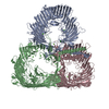
|
|---|---|
| 1 |
|
-Components
| #1: Protein | Mass: 123835.367 Da / Num. of mol.: 3 / Source method: isolated from a natural source / Source: (natural) Deinococcus radiodurans (radioresistant)Strain: ATCC 13939 / DSM 20539 / JCM 16871 / LMG 4051 / NBRC 15346 / NCIMB 9279 / R1 / VKM B-1422 References: UniProt: Q9RRB6 #2: Chemical | ChemComp-CA /   Mass: 40.078 Da / Num. of mol.: 18 / Source method: obtained synthetically / Formula: Ca Mass: 40.078 Da / Num. of mol.: 18 / Source method: obtained synthetically / Formula: CaHas ligand of interest | N | Has protein modification | Y | |
|---|
-Experimental details
-Experiment
| Experiment | Method: ELECTRON MICROSCOPY |
|---|---|
| EM experiment | Aggregation state: PARTICLE / 3D reconstruction method: single particle reconstruction |
- Sample preparation
| Component | Name: Structure of trimeric SlpA protein / Type: COMPLEX / Details: Structure of trimeric SlpA protein / Entity ID: #1 / Source: NATURAL | ||||||||||||||||||||
|---|---|---|---|---|---|---|---|---|---|---|---|---|---|---|---|---|---|---|---|---|---|
| Molecular weight | Experimental value: NO | ||||||||||||||||||||
| Source (natural) | Organism: Deinococcus radiodurans (radioresistant) / Strain: BAA-816 | ||||||||||||||||||||
| Buffer solution | pH: 7.5 Details: Buffer solutions were prepared fresh from sterile filtered concentrated stocksolutions. Solutions were filtered through a 0.22 um filter to avoid microbial contamination and degassed using a ...Details: Buffer solutions were prepared fresh from sterile filtered concentrated stocksolutions. Solutions were filtered through a 0.22 um filter to avoid microbial contamination and degassed using a vacuum fold pump. The pH of the HEPES stock solution was adjusted with sodium hydroxide at 4 deg C. | ||||||||||||||||||||
| Buffer component |
| ||||||||||||||||||||
| Specimen | Conc.: 4.45 mg/ml / Embedding applied: NO / Shadowing applied: NO / Staining applied: NO / Vitrification applied: YES Details: Purified SlpA protein after size-exclusion chromatography | ||||||||||||||||||||
| Specimen support | Details: 20 seconds, 15 mA / Grid material: COPPER/RHODIUM / Grid mesh size: 200 divisions/in. / Grid type: Quantifoil R2/2 | ||||||||||||||||||||
| Vitrification | Instrument: FEI VITROBOT MARK IV / Cryogen name: ETHANE / Humidity: 100 % / Chamber temperature: 283.15 K Details: Vitrobot options: Blot time 4.5 seconds, Blot force -10,1, Wait time 10 seconds, Drain time 0.5 seconds |
- Electron microscopy imaging
| Experimental equipment |  Model: Titan Krios / Image courtesy: FEI Company |
|---|---|
| Microscopy | Model: TFS KRIOS Details: EPU software with faster acquisition mode AFIS (Aberration Free Image Shift). |
| Electron gun | Electron source:  FIELD EMISSION GUN / Accelerating voltage: 300 kV / Illumination mode: FLOOD BEAM FIELD EMISSION GUN / Accelerating voltage: 300 kV / Illumination mode: FLOOD BEAM |
| Electron lens | Mode: BRIGHT FIELD / Nominal magnification: 81000 X / Calibrated magnification: 81000 X / Nominal defocus max: 4000 nm / Nominal defocus min: 1000 nm / Calibrated defocus min: 1000 nm / Calibrated defocus max: 4000 nm / Cs: 2.7 mm / C2 aperture diameter: 50 µm / Alignment procedure: ZEMLIN TABLEAU |
| Specimen holder | Cryogen: NITROGEN / Specimen holder model: FEI TITAN KRIOS AUTOGRID HOLDER / Temperature (max): 70 K / Temperature (min): 70 K |
| Image recording | Average exposure time: 4.8 sec. / Electron dose: 47.909 e/Å2 / Film or detector model: GATAN K3 BIOQUANTUM (6k x 4k) / Num. of grids imaged: 1 / Num. of real images: 2294 |
| EM imaging optics | Energyfilter name: GIF Bioquantum / Energyfilter slit width: 20 eV |
| Image scans | Width: 5760 / Height: 4092 |
- Processing
| Software | Name: PHENIX / Version: 1.19.1_4122: / Classification: refinement | ||||||||||||||||||||||||||||||||||||||||||||||||||
|---|---|---|---|---|---|---|---|---|---|---|---|---|---|---|---|---|---|---|---|---|---|---|---|---|---|---|---|---|---|---|---|---|---|---|---|---|---|---|---|---|---|---|---|---|---|---|---|---|---|---|---|
| EM software |
| ||||||||||||||||||||||||||||||||||||||||||||||||||
| Image processing | Details: Movies were clustered into optics groups based on the XML meta-data of the data-collection software EPU (ThermoFisher) using a k-means algorithm implemented in EPU_group_AFIS (https://github. ...Details: Movies were clustered into optics groups based on the XML meta-data of the data-collection software EPU (ThermoFisher) using a k-means algorithm implemented in EPU_group_AFIS (https://github.com/DustinMorado/EPU_group_AFIS). Imported movies were motion-corrected, dose weighted, and Fourier cropped (2x) with MotionCor2 (Zheng et al., 2017) implemented in RELION3.1 (Zivanov et al., 2018). Contrast transfer functions (CTFs) of the resulting motion-corrected micrographs were estimated using CTFFIND4 (Rohou and Grigorieff, 2015). | ||||||||||||||||||||||||||||||||||||||||||||||||||
| CTF correction | Details: RELION refinement with in-built CTF correction. The function is similar to a Wiener filter, so amplitude correction included. Type: PHASE FLIPPING AND AMPLITUDE CORRECTION | ||||||||||||||||||||||||||||||||||||||||||||||||||
| Particle selection | Num. of particles selected: 223878 Details: Initially, micrographs were denoised using TOPAZ (73) using the UNET neural network and 2893 particles were manually picked. Particle coordinates were used to train TOPAZ picker (74) in 5 ...Details: Initially, micrographs were denoised using TOPAZ (73) using the UNET neural network and 2893 particles were manually picked. Particle coordinates were used to train TOPAZ picker (74) in 5 times downsampled micrographs with the neural network architecture ResNet8 and picked particles were extracted in 4 times downsampled 128 x 128 boxes and classified using reference-free 2D classification inside RELION3.1. | ||||||||||||||||||||||||||||||||||||||||||||||||||
| Symmetry | Point symmetry: C3 (3 fold cyclic) | ||||||||||||||||||||||||||||||||||||||||||||||||||
| 3D reconstruction | Resolution: 3.25 Å / Resolution method: FSC 0.143 CUT-OFF / Num. of particles: 122412 / Algorithm: FOURIER SPACE / Num. of class averages: 1 / Symmetry type: POINT | ||||||||||||||||||||||||||||||||||||||||||||||||||
| Atomic model building | B value: 51.67 / Protocol: AB INITIO MODEL / Space: REAL / Target criteria: Best Fit | ||||||||||||||||||||||||||||||||||||||||||||||||||
| Refine LS restraints |
|
