 Movie
Movie Controller


+ Open data

- Basic information
| Entry |  | ||||||||||||
|---|---|---|---|---|---|---|---|---|---|---|---|---|---|
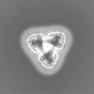
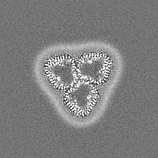
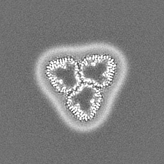
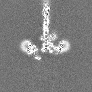
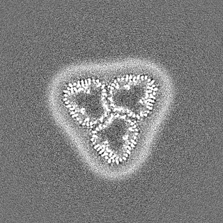
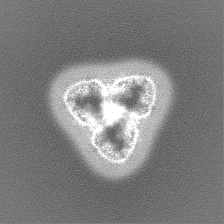
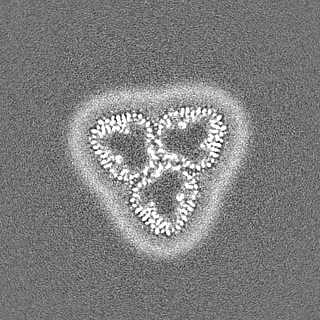
| Title | Structure of trimeric SlpA outer membrane protein | ||||||||||||
 Map data Map data | RELION PostProcessed map with B-factor sharpening | ||||||||||||
 Sample Sample |
| ||||||||||||
 Keywords Keywords | SlpA protein / STRUCTURAL PROTEIN | ||||||||||||
| Function / homology |  Function and homology information Function and homology informationporin activity / pore complex / monoatomic ion transport / cell outer membrane / lipid binding Similarity search - Function | ||||||||||||
| Biological species |  Deinococcus radiodurans (radioresistant) Deinococcus radiodurans (radioresistant) | ||||||||||||
| Method | single particle reconstruction / cryo EM / Resolution: 3.25 Å | ||||||||||||
 Authors Authors | von Kuegelgen A / Bharat TAM | ||||||||||||
| Funding support |  United Kingdom, 3 items United Kingdom, 3 items
| ||||||||||||
 Citation Citation | Journal: Proc Natl Acad Sci U S A / Year: 2022 Title: A multidomain connector links the outer membrane and cell wall in phylogenetically deep-branching bacteria. Authors: Andriko von Kügelgen / Sofie van Dorst / Vikram Alva / Tanmay A M Bharat /  Abstract: is a phylogenetically deep-branching extremophilic bacterium that is remarkably tolerant to numerous environmental stresses, including large doses of ultraviolet (UV) radiation and extreme ... is a phylogenetically deep-branching extremophilic bacterium that is remarkably tolerant to numerous environmental stresses, including large doses of ultraviolet (UV) radiation and extreme temperatures. It can even survive in outer space for several years. This endurance of has been partly ascribed to its atypical cell envelope comprising an inner membrane, a large periplasmic space with a thick peptidoglycan (PG) layer, and an outer membrane (OM) covered by a surface layer (S-layer). Despite intense research, molecular principles governing envelope organization and OM stabilization are unclear in and related bacteria. Here, we report a electron cryomicroscopy (cryo-EM) structure of the abundant OM protein SlpA, showing how its C-terminal segment forms homotrimers of 30-stranded β-barrels in the OM, whereas its N-terminal segment forms long, homotrimeric coiled coils linking the OM to the PG layer via S-layer homology (SLH) domains. Furthermore, using protein structure prediction and sequence-based bioinformatic analysis, we show that SlpA-like putative OM-PG connector proteins are widespread in phylogenetically deep-branching Gram-negative bacteria. Finally, combining our atomic structures with fluorescence and electron microscopy of cell envelopes of wild-type and mutant bacterial strains, we report a model for the cell surface of . Our results will have important implications for understanding the cell surface organization and hyperstability of and related bacteria and the evolutionary transition between Gram-negative and Gram-positive bacteria. | ||||||||||||
| History |
|
- Structure visualization
| Supplemental images |
|---|

- Downloads & links
-EMDB archive
| Map data | emd_15378.map.gz | 116.2 MB | EMDB map data format | |
|---|---|---|---|---|
| Header (meta data) | emd-15378-v30.xmlemd-15378.xml | 25 KB 25 KB | Display Display | EMDB header |
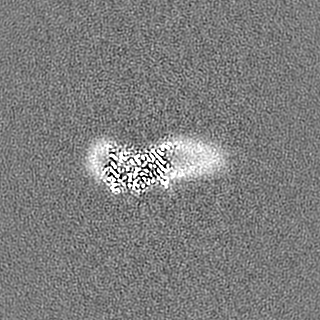
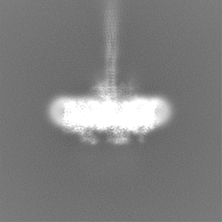
| Images | 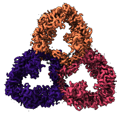 emd_15378.png emd_15378.png | 184.4 KB | ||
| Masks | emd_15378_msk_1.map | 125 MB | Mask map | |
| Filedesc metadata | emd-15378.cif.gz | 7.8 KB | ||
| Others | emd_15378_additional_1.map.gzemd_15378_half_map_1.map.gzemd_15378_half_map_2.map.gz | 115.7 MB 116 MB 116 MB | ||
| Archive directory |  http://ftp.pdbj.org/pub/emdb/structures/EMD-15378ftp://ftp.pdbj.org/pub/emdb/structures/EMD-15378 http://ftp.pdbj.org/pub/emdb/structures/EMD-15378ftp://ftp.pdbj.org/pub/emdb/structures/EMD-15378 | HTTPS FTP |
-Related structure data
| Related structure data |  8ae1MC M: atomic model generated by this map C: citing same article ( |
|---|---|
| Similar structure data |
-Links
| EMDB pages | EMDB (EBI/PDBe) / EMDataResource |
|---|
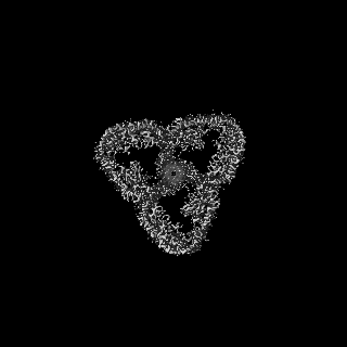
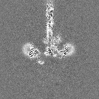
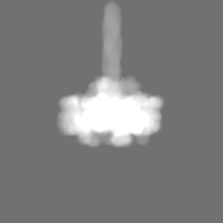
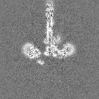
-Map
| File | Download / File: emd_15378.map.gz / Format: CCP4 / Size: 125 MB / Type: IMAGE STORED AS FLOATING POINT NUMBER (4 BYTES) | ||||||||||||||||||||||||||||||||||||
|---|---|---|---|---|---|---|---|---|---|---|---|---|---|---|---|---|---|---|---|---|---|---|---|---|---|---|---|---|---|---|---|---|---|---|---|---|---|
| Annotation | RELION PostProcessed map with B-factor sharpening | ||||||||||||||||||||||||||||||||||||
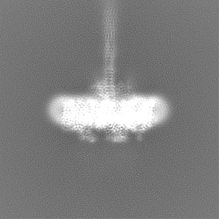
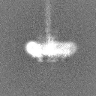
| Projections & slices | Image control
Images are generated by Spider. | ||||||||||||||||||||||||||||||||||||
| Voxel size | X=Y=Z: 1.092 Å | ||||||||||||||||||||||||||||||||||||
| Density |
| ||||||||||||||||||||||||||||||||||||
| Symmetry | Space group: 1 | ||||||||||||||||||||||||||||||||||||
| Details | EMDB XML:
|
 Z (Sec.)
 Y (Row.)
 X (Col.)


-Supplemental data
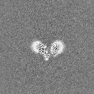
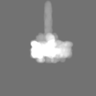
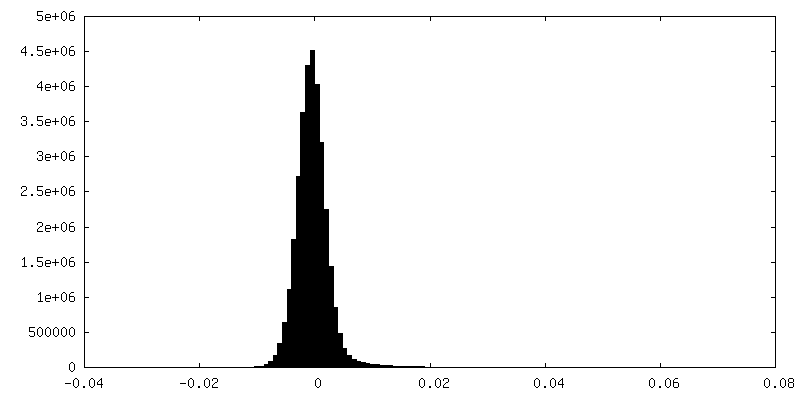
-Mask #1
| File | emd_15378_msk_1.map | ||||||||||||
|---|---|---|---|---|---|---|---|---|---|---|---|---|---|
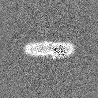
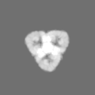
| Projections & Slices |
| ||||||||||||
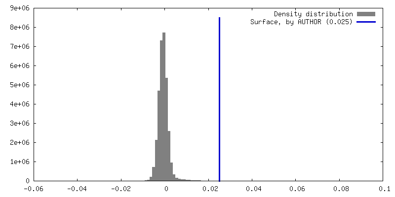
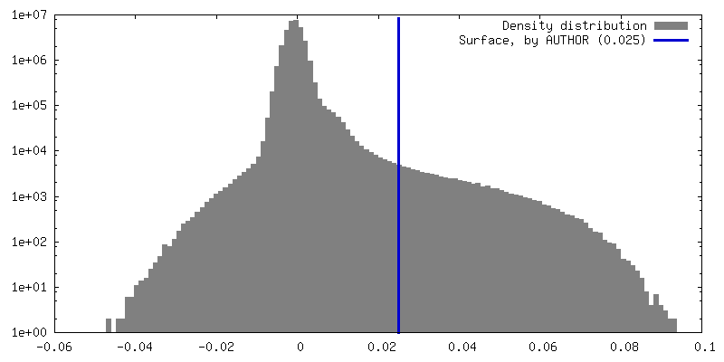
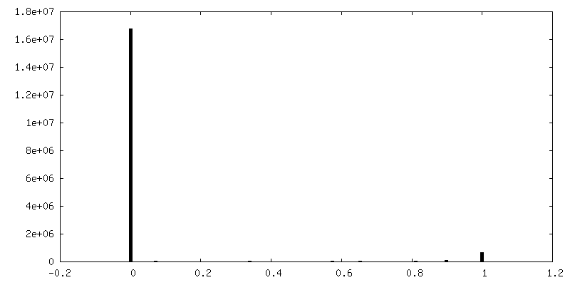
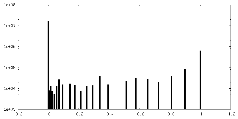
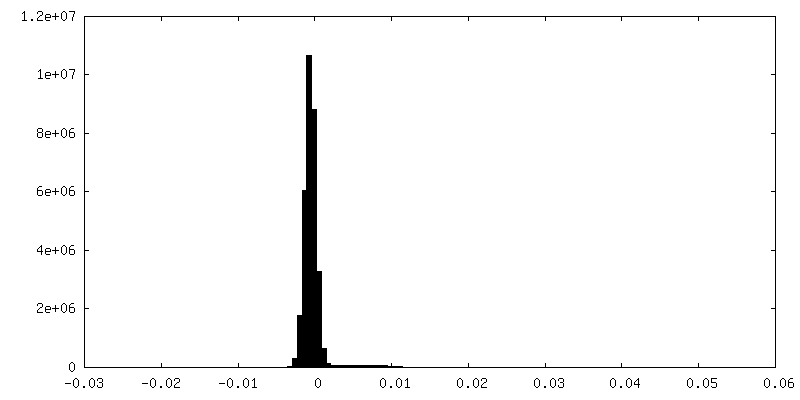
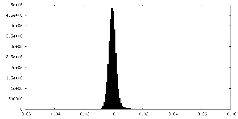
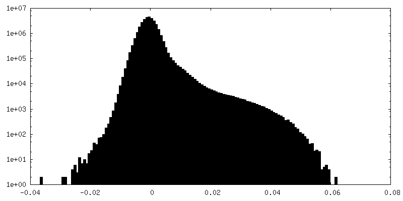
| Density Histograms |


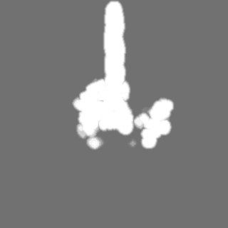
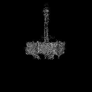
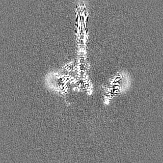
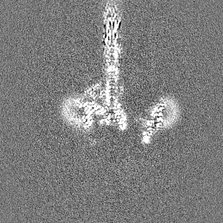
-Additional map: Full map RELION without B-factor sharpening
| File | emd_15378_additional_1.map | ||||||||||||
|---|---|---|---|---|---|---|---|---|---|---|---|---|---|
| Annotation | Full map RELION without B-factor sharpening | ||||||||||||
| Projections & Slices |
| ||||||||||||
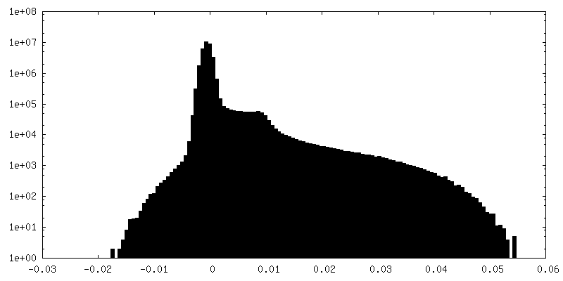
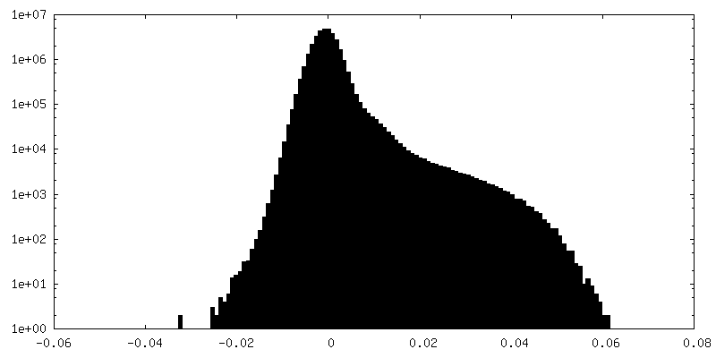
| Density Histograms |


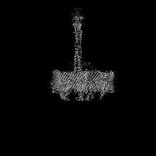
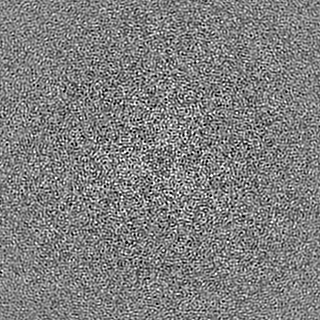
-Half map: RELION Half map 1
| File | emd_15378_half_map_1.map | ||||||||||||
|---|---|---|---|---|---|---|---|---|---|---|---|---|---|
| Annotation | RELION Half map 1 | ||||||||||||
| Projections & Slices |
| ||||||||||||
| Density Histograms |


-Half map: RELION Half map 1
| File | emd_15378_half_map_2.map | ||||||||||||
|---|---|---|---|---|---|---|---|---|---|---|---|---|---|
| Annotation | RELION Half map 1 | ||||||||||||
| Projections & Slices |
| ||||||||||||
| Density Histograms |

- Sample components
-Entire : Structure of trimeric SlpA protein
| Entire | Name: Structure of trimeric SlpA protein |
|---|---|
| Components |
|
-Supramolecule #1: Structure of trimeric SlpA protein
| Supramolecule | Name: Structure of trimeric SlpA protein / type: complex / ID: 1 / Parent: 0 / Macromolecule list: #1 / Details: Structure of trimeric SlpA protein |
|---|---|
| Source (natural) | Organism: Deinococcus radiodurans (radioresistant) / Strain: BAA-816 |
-Macromolecule #1: S-layer protein SlpA
| Macromolecule | Name: S-layer protein SlpA / type: protein_or_peptide / ID: 1 / Number of copies: 3 / Enantiomer: LEVO |
|---|---|
| Source (natural) | Organism: Deinococcus radiodurans (radioresistant) Strain: ATCC 13939 / DSM 20539 / JCM 16871 / LMG 4051 / NBRC 15346 / NCIMB 9279 / R1 / VKM B-1422 |
| Molecular weight | Theoretical: 123.835367 KDa |
| Sequence | String: MKKSLIALTT ALSFGLAAAQ TAAPVSAPQV PALTDVPAGH WAKDAIDRLV SRGVILGYPD GTFRGTQNLT RYEAAIIIAR LLDQMRDGE TPAGMTAEDM TALQNAIQEL AADLAALGVR VSDLEANAVS KDDFARLEAR IEEVAAAGGE QGATEALQGQ I DDLTARVD ...String: MKKSLIALTT ALSFGLAAAQ TAAPVSAPQV PALTDVPAGH WAKDAIDRLV SRGVILGYPD GTFRGTQNLT RYEAAIIIAR LLDQMRDGE TPAGMTAEDM TALQNAIQEL AADLAALGVR VSDLEANAVS KDDFARLEAR IEEVAAAGGE QGATEALQGQ I DDLTARVD EYDALRADVD DNASSIAALN DLTVLLNQDI LDLQDRVSAV EAAQADFVQR SDFDALGGRV TTVETRVETV NN SLTGRIA ALERNAFSVK PSLTIGYSVS RTSRNFDVDR LFPLNADGTV ANNAFTSGGI DTDTGAQRRD FGDFGNASDP VVA GAAGLY GFADGVSYTV YFTDGSTATF DGLNPADYKV PTGKVIDTTK GRNGFGFNNL ARYKEGSTDI GISLGFDTSG QFSQ VTSGT GGSLFSTAGR LQVNQIDLNF GLVTGLPSDA YVDTNGNGKK DDGEATGRGT YLGSGGTAAI LRDPAGNVYR PVFFR FKNA TTQFSVGNNP VIVTLGQQQK FYFSDYVFDN NYDGRGDGFT VTVDGSNVPV IGAWKPQIKG VYGSRSGLDG TAEAGY GVY YRGVRAQITP VGTLTAGIHY AQEGRDMFGA AQNTTSTPSD VTTYGADLHG KAFGVELHSE YATSRVRPNT ANAAVQT SN AFYARVATRK DNLAFDLNTP AAKFGNDTFG VSLYDLNYRK IDAGYNNVAG ISEYGYGSYS RTSAQNIAYN PDTGVTAP F ANLDRQAYTD ANNDGTSDRN ADGTVVATNT KIGQMGFGVK AAANLGPVAI GGYYDTSTGA NGDNANRMTE AGGSAKVAY SIFSLRGTYN TLDSNRPQIY RDAAGTQIIG DAKVRRYAVQ ADVTPGLGLF VGAYYRDVNV NGVRSTTDRG LLGRGYLASS FEPGVGNNA YRTGLRCADN NFGTGTRDID GVGGVLNPAV NLDQSRTATC FTSYGVEAGH AGDNANALVK DLFFRVGYSR V YVPTTATA TTGDFSGSVT YGDARYDRKV GVANVRLAGS FSTTNTQLDS RPAGTRGAVG LIVRTDPLEN VPFRPQFNGQ VG YYTADNR VAAGNYNANA TKYGAGVVLN DFLLPQTKIG VRYDGYMAQN RQYTPFDGDG TQGYFSDANN NRRTNLNGVY VEG AYQDLI FSYGTYTLSQ KDLNGVEYGS GINNGQPARG QTFKISYKVN F UniProtKB: Outer membrane protein SlpA |
-Macromolecule #2: CALCIUM ION
| Macromolecule | Name: CALCIUM ION / type: ligand / ID: 2 / Number of copies: 18 / Formula: CA |
|---|---|
| Molecular weight | Theoretical: 40.078 Da |
-Experimental details
-Structure determination
| Method | cryo EM |
|---|---|
 Processing Processing | single particle reconstruction |
| Aggregation state | particle |
-Sample preparation
| Concentration | 4.45 mg/mL | ||||||||||||
|---|---|---|---|---|---|---|---|---|---|---|---|---|---|
| Buffer | pH: 7.5 Component:
Details: Buffer solutions were prepared fresh from sterile filtered concentrated stocksolutions. Solutions were filtered through a 0.22 um filter to avoid microbial contamination and degassed using a ...Details: Buffer solutions were prepared fresh from sterile filtered concentrated stocksolutions. Solutions were filtered through a 0.22 um filter to avoid microbial contamination and degassed using a vacuum fold pump. The pH of the HEPES stock solution was adjusted with sodium hydroxide at 4 deg C. | ||||||||||||
| Grid | Model: Quantifoil R2/2 / Material: COPPER/RHODIUM / Mesh: 200 / Support film - Material: CARBON / Support film - topology: HOLEY / Pretreatment - Type: GLOW DISCHARGE / Pretreatment - Time: 20 sec. / Pretreatment - Atmosphere: AIR / Details: 20 seconds, 15 mA | ||||||||||||
| Vitrification | Cryogen name: ETHANE / Chamber humidity: 100 % / Chamber temperature: 283.15 K / Instrument: FEI VITROBOT MARK IV Details: Vitrobot options: Blot time 4.5 seconds, Blot force -10,1, Wait time 10 seconds, Drain time 0.5 seconds. | ||||||||||||
| Details | Purified SlpA protein after size-exclusion chromatography |
- Electron microscopy
| Microscope | TFS KRIOS |
|---|---|
| Temperature | Min: 70.0 K / Max: 70.0 K |
| Specialist optics | Energy filter - Name: GIF Bioquantum / Energy filter - Slit width: 20 eV |
| Details | EPU software with faster acquisition mode AFIS (Aberration Free Image Shift). |
| Image recording | Film or detector model: GATAN K3 BIOQUANTUM (6k x 4k) / Digitization - Dimensions - Width: 5760 pixel / Digitization - Dimensions - Height: 4092 pixel / Number grids imaged: 1 / Number real images: 2294 / Average exposure time: 4.8 sec. / Average electron dose: 47.909 e/Å2 |
| Electron beam | Acceleration voltage: 300 kV / Electron source:  FIELD EMISSION GUN FIELD EMISSION GUN |
| Electron optics | C2 aperture diameter: 50.0 µm / Calibrated defocus max: 4.0 µm / Calibrated defocus min: 1.0 µm / Calibrated magnification: 81000 / Illumination mode: FLOOD BEAM / Imaging mode: BRIGHT FIELD / Cs: 2.7 mm / Nominal defocus max: 4.0 µm / Nominal defocus min: 1.0 µm / Nominal magnification: 81000 |
| Sample stage | Specimen holder model: FEI TITAN KRIOS AUTOGRID HOLDER / Cooling holder cryogen: NITROGEN |
| Experimental equipment |  Model: Titan Krios / Image courtesy: FEI Company |
+Image processing
-Atomic model buiding 1
| Refinement | Space: REAL / Protocol: AB INITIO MODEL / Overall B value: 51.67 / Target criteria: Best Fit |
|---|---|
| Output model | PDB-8ae1: |
