 Movie
Movie Controller


+ Open data

- Basic information
| Entry | Database: PDB / ID: 7zj2 | ||||||
|---|---|---|---|---|---|---|---|
| Title | Amyloid fibril (in vitro) from full-length hnRNPA1 protein | ||||||
 Components Components | Isoform A1-A of Heterogeneous nuclear ribonucleoprotein A1 | ||||||
 Keywords Keywords | NUCLEAR PROTEIN / Amyloidosis / Misfolding disease / Inflammation / Prion / Protein fibril | ||||||
| Function / homology |  Function and homology information Function and homology informationcellular response to sodium arsenite / SARS-CoV-1-host interactions / import into nucleus / telomeric repeat-containing RNA binding / G-rich strand telomeric DNA binding / pre-mRNA binding / nuclear export / RNA export from nucleus / miRNA binding / FGFR2 alternative splicing ...cellular response to sodium arsenite / SARS-CoV-1-host interactions / import into nucleus / telomeric repeat-containing RNA binding / G-rich strand telomeric DNA binding / pre-mRNA binding / nuclear export / RNA export from nucleus / miRNA binding / FGFR2 alternative splicing / regulation of alternative mRNA splicing, via spliceosome / SARS-CoV-1 modulates host translation machinery / regulation of RNA splicing / negative regulation of telomere maintenance via telomerase / Processing of Capped Intron-Containing Pre-mRNA / mRNA transport / cellular response to glucose starvation / positive regulation of telomere maintenance via telomerase / catalytic step 2 spliceosome / mRNA Splicing - Major Pathway / mRNA 3'-UTR binding / spliceosomal complex / mRNA splicing, via spliceosome / single-stranded DNA binding / single-stranded RNA binding / ribonucleoprotein complex / protein domain specific binding / DNA binding / RNA binding / extracellular exosome / nucleoplasm / identical protein binding / membrane / nucleus / cytosol / cytoplasm Similarity search - Function | ||||||
| Biological species |  Homo sapiens (human) Homo sapiens (human) | ||||||
| Method | ELECTRON MICROSCOPY / helical reconstruction / cryo EM / Resolution: 3.32 Å | ||||||
 Authors Authors | Sharma, K. / Banerjee, S. / Schmidt, M. / Faendrich, M. | ||||||
| Funding support | 1items
| ||||||
 Citation Citation | Journal: J Mol Biol / Year: 2023 Title: Cryo-EM Structure of the Full-length hnRNPA1 Amyloid Fibril. Authors: Kartikay Sharma / Sambhasan Banerjee / Dilan Savran / Cedric Rajes / Sebastian Wiese / Amandeep Girdhar / Nadine Schwierz / Christopher Lee / James Shorter / Matthias Schmidt / Lin Guo / Marcus Fändrich /   Abstract: Heterogeneous nuclear ribonucleoprotein A1 (hnRNPA1) is a multifunctional RNA-binding protein that is associated with neurodegenerative diseases, such as amyotrophic lateral sclerosis and multisystem ...Heterogeneous nuclear ribonucleoprotein A1 (hnRNPA1) is a multifunctional RNA-binding protein that is associated with neurodegenerative diseases, such as amyotrophic lateral sclerosis and multisystem proteinopathy. In this study, we have used cryo-electron microscopy to investigate the three-dimensional structure of amyloid fibrils from full-length hnRNPA1 protein. We find that the fibril core is formed by a 45-residue segment of the prion-like low-complexity domain of the protein, whereas the remaining parts of the protein (275 residues) form a fuzzy coat around the fibril core. The fibril consists of two fibril protein stacks that are arranged into a pseudo-2 screw symmetry. The ordered core harbors several of the positions that are known to be affected by disease-associated mutations, but does not encompass the most aggregation-prone segments of the protein. These data indicate that the structures of amyloid fibrils from full-length proteins may be more complex than anticipated by current theories on protein misfolding. | ||||||
| History |
|
- Structure visualization
| Structure viewer | Molecule: MolmilJmol/JSmol |
|---|
- Downloads & links
-Download
| PDBx/mmCIF format | 7zj2.cif.gz | 127.1 KB | Display | PDBx/mmCIF format |
|---|---|---|---|---|
| PDB format | pdb7zj2.ent.gz | 79.9 KB | Display | PDB format |
| PDBx/mmJSON format | 7zj2.json.gz | Tree view | PDBx/mmJSON format | |
| Others |  Other downloads Other downloads |
-Validation report
| Arichive directory | https://data.pdbj.org/pub/pdb/validation_reports/zj/7zj2ftp://data.pdbj.org/pub/pdb/validation_reports/zj/7zj2 | HTTPS FTP |
|---|
-Related structure data
| Related structure data | 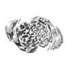 14739MC M: map data used to model this data C: citing same article ( |
|---|---|
| Similar structure data |
-Links
 PDBj
PDBj


- Assembly
| Deposited unit | 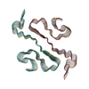
|
|---|---|
| 1 |
|
-Components
| #1: Protein | Mass: 34246.227 Da / Num. of mol.: 12 Source method: isolated from a genetically manipulated source Source: (gene. exp.) Homo sapiens (human) / Gene: HNRNPA1, HNRPA1 / Production host:  |
|---|
-Experimental details
-Experiment
| Experiment | Method: ELECTRON MICROSCOPY |
|---|---|
| EM experiment | Aggregation state: HELICAL ARRAY / 3D reconstruction method: helical reconstruction |
- Sample preparation
| Component | Name: Amyloid fibril (in vitro) from full-length hnRNPA1 protein Type: COMPLEX / Details: In vitro full-length hnRNPA1 amyloid fibril / Entity ID: all / Source: RECOMBINANT |
|---|---|
| Molecular weight | Experimental value: NO |
| Source (natural) | Organism: Homo sapiens (human) |
| Source (recombinant) | Organism: |
| Buffer solution | pH: 7.4 |
| Specimen | Embedding applied: NO / Shadowing applied: NO / Staining applied: NO / Vitrification applied: YES |
| Specimen support | Grid material: COPPER / Grid mesh size: 400 divisions/in. / Grid type: C-flat-1.2/1.3 |
| Vitrification | Instrument: FEI VITROBOT MARK III / Cryogen name: ETHANE / Humidity: 95 % |
- Electron microscopy imaging
| Experimental equipment |  Model: Titan Krios / Image courtesy: FEI Company |
|---|---|
| Microscopy | Model: FEI TITAN KRIOS |
| Electron gun | Electron source:  FIELD EMISSION GUN / Accelerating voltage: 300 kV / Illumination mode: FLOOD BEAM FIELD EMISSION GUN / Accelerating voltage: 300 kV / Illumination mode: FLOOD BEAM |
| Electron lens | Mode: BRIGHT FIELD / Nominal defocus max: 2000 nm / Nominal defocus min: 1000 nm / Cs: 2.7 mm |
| Specimen holder | Cryogen: NITROGEN |
| Image recording | Average exposure time: 10 sec. / Electron dose: 42.64 e/Å2 / Detector mode: COUNTING / Film or detector model: GATAN K2 SUMMIT (4k x 4k) / Num. of real images: 2624 |
| Image scans | Movie frames/image: 40 |
- Processing
| EM software |
| ||||||||||||||||||||||||||||||||
|---|---|---|---|---|---|---|---|---|---|---|---|---|---|---|---|---|---|---|---|---|---|---|---|---|---|---|---|---|---|---|---|---|---|
| CTF correction | Type: PHASE FLIPPING AND AMPLITUDE CORRECTION | ||||||||||||||||||||||||||||||||
| Helical symmerty | Angular rotation/subunit: 179.05 ° / Axial rise/subunit: 2.37 Å / Axial symmetry: C1 | ||||||||||||||||||||||||||||||||
| Particle selection | Num. of particles selected: 184301 | ||||||||||||||||||||||||||||||||
| 3D reconstruction | Resolution: 3.32 Å / Resolution method: FSC 0.143 CUT-OFF / Num. of particles: 54408 / Symmetry type: HELICAL | ||||||||||||||||||||||||||||||||
| Atomic model building | Protocol: BACKBONE TRACE / Space: REAL / Target criteria: correlation coefficient | ||||||||||||||||||||||||||||||||
| Atomic model building | PDB-ID: 7BX7 Accession code: 7BX7 / Source name: PDB / Type: experimental model |

