+ データを開く
- 基本情報
基本情報
| 登録情報 | データベース: PDB / ID: 7ypx | ||||||
|---|---|---|---|---|---|---|---|
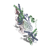
| タイトル | Cyanophage Pam3 fiber | ||||||
 要素 要素 |
| ||||||
 キーワード キーワード | VIRAL PROTEIN / fiber / VIRUS | ||||||
| 生物種 |  uncultured cyanophage (ファージ) uncultured cyanophage (ファージ) | ||||||
| 手法 | 電子顕微鏡法 / 単粒子再構成法 / クライオ電子顕微鏡法 / 解像度: 3.12 Å | ||||||
 データ登録者 データ登録者 | Wei, Z.L. / Jiang, Y.L. / Zhou, C.Z. | ||||||
| 資金援助 |  中国, 1件 中国, 1件
| ||||||
 引用 引用 |  ジャーナル: Viruses / 年: 2022 ジャーナル: Viruses / 年: 2022タイトル: Structural Insights into the Chaperone-Assisted Assembly of a Simplified Tail Fiber of the Myocyanophage Pam3. 著者: Zi-Lu Wei / Feng Yang / Bo Li / Pu Hou / Wen-Wen Kong / Jie Wang / Yuxing Chen / Yong-Liang Jiang / Cong-Zhao Zhou /  要旨: At the first step of phage infection, the receptor-binding proteins (RBPs) such as tail fibers are responsible for recognizing specific host surface receptors. The proper folding and assembly of tail ...At the first step of phage infection, the receptor-binding proteins (RBPs) such as tail fibers are responsible for recognizing specific host surface receptors. The proper folding and assembly of tail fibers usually requires a chaperone encoded by the phage genome. Despite extensive studies on phage structures, the molecular mechanism of phage tail fiber assembly remains largely unknown. Here, using a minimal myocyanophage, termed Pam3, isolated from Lake Chaohu, we demonstrate that the chaperone gp25 forms a stable complex with the tail fiber gp24 at a stoichiometry of 3:3. The 3.1-Å cryo-electron microscopy structure of this complex revealed an elongated structure with the gp25 trimer embracing the distal moieties of gp24 trimer at the center. Each gp24 subunit consists of three domains: the N-terminal α-helical domain required for docking to the baseplate, the tumor necrosis factor (TNF)-like and glycine-rich domains responsible for recognizing the host receptor. Each gp25 subunit consists of two domains: a non-conserved N-terminal β-sandwich domain that binds to the TNF-like and glycine-rich domains of the fiber, and a C-terminal α-helical domain that mediates trimerization/assembly of the fiber. Structural analysis enabled us to propose the assembly mechanism of phage tail fibers, in which the chaperone first protects the intertwined and repetitive distal moiety of each fiber subunit, further ensures the proper folding of these highly plastic structural elements, and eventually enables the formation of the trimeric fiber. These findings provide the structural basis for the design and engineering of phage fibers for biotechnological applications. | ||||||
| 履歴 |
|
- 構造の表示
構造の表示
| 構造ビューア | 分子:  Molmil Molmil Jmol/JSmol Jmol/JSmol |
|---|
- ダウンロードとリンク
- ダウンロード
| PDBx/mmCIF形式 |  7ypx.cif.gz 7ypx.cif.gz | 198.8 KB | 表示 |  PDBx/mmCIF形式 PDBx/mmCIF形式 |
|---|---|---|---|---|
| PDB形式 |  pdb7ypx.ent.gz pdb7ypx.ent.gz | 161.3 KB | 表示 |  PDB形式 PDB形式 |
| PDBx/mmJSON形式 |  7ypx.json.gz 7ypx.json.gz | ツリー表示 |  PDBx/mmJSON形式 PDBx/mmJSON形式 | |
| その他 |  その他のダウンロード その他のダウンロード |
-検証レポート
| 文書・要旨 |  7ypx_validation.pdf.gz 7ypx_validation.pdf.gz | 1004.8 KB | 表示 |  wwPDB検証レポート wwPDB検証レポート |
|---|---|---|---|---|
| 文書・詳細版 |  7ypx_full_validation.pdf.gz 7ypx_full_validation.pdf.gz | 1015.5 KB | 表示 | |
| XML形式データ |  7ypx_validation.xml.gz 7ypx_validation.xml.gz | 41.2 KB | 表示 | |
| CIF形式データ |  7ypx_validation.cif.gz 7ypx_validation.cif.gz | 62 KB | 表示 | |
| アーカイブディレクトリ |  https://data.pdbj.org/pub/pdb/validation_reports/yp/7ypx https://data.pdbj.org/pub/pdb/validation_reports/yp/7ypx ftp://data.pdbj.org/pub/pdb/validation_reports/yp/7ypx ftp://data.pdbj.org/pub/pdb/validation_reports/yp/7ypx | HTTPS FTP |
-関連構造データ
- リンク
リンク
- 集合体
集合体
| 登録構造単位 | 
|
|---|---|
| 1 |
|
- 要素
要素
| #1: タンパク質 | 分子量: 28383.873 Da / 分子数: 3 / 由来タイプ: 組換発現 詳細: The source organism is the myocyanophage Pam3 isolated from the Lake Chaohu, China. 由来: (組換発現)  uncultured cyanophage (ファージ) uncultured cyanophage (ファージ)発現宿主:  #2: タンパク質 | 分子量: 17457.660 Da / 分子数: 3 / 由来タイプ: 組換発現 詳細: The source organism is the myocyanophage Pam3 isolated from the Lake Chaohu, China. 由来: (組換発現)  uncultured cyanophage (ファージ) uncultured cyanophage (ファージ)発現宿主:  |
|---|
-実験情報
-実験
| 実験 | 手法: 電子顕微鏡法 |
|---|---|
| EM実験 | 試料の集合状態: PARTICLE / 3次元再構成法: 単粒子再構成法 |
- 試料調製
試料調製
| 構成要素 | 名称: Pam3 tail fiber / タイプ: COMPLEX / Entity ID: all / 由来: RECOMBINANT |
|---|---|
| 由来(天然) | 生物種:  uncultured cyanophage (ファージ) uncultured cyanophage (ファージ) |
| 由来(組換発現) | 生物種:  |
| 緩衝液 | pH: 7 |
| 試料 | 包埋: NO / シャドウイング: NO / 染色: NO / 凍結: YES |
| 急速凍結 | 凍結剤: ETHANE / 湿度: 100 % / 凍結前の試料温度: 300 K |
- 電子顕微鏡撮影
| 実験機器 |  モデル: Titan Krios / 画像提供: FEI Company |
|---|---|
| 顕微鏡 | モデル: FEI TITAN KRIOS |
| 電子銃 | 電子線源:  FIELD EMISSION GUN / 加速電圧: 300 kV / 照射モード: SPOT SCAN FIELD EMISSION GUN / 加速電圧: 300 kV / 照射モード: SPOT SCAN |
| 電子レンズ | モード: BRIGHT FIELD / 最大 デフォーカス(公称値): 2500 nm / 最小 デフォーカス(公称値): 1500 nm |
| 撮影 | 電子線照射量: 55 e/Å2 / フィルム・検出器のモデル: GATAN K3 (6k x 4k) |
- 解析
解析
| ソフトウェア | 名称: PHENIX / バージョン: 1.19.2_4158: / 分類: 精密化 | ||||||||||||||||||||||||
|---|---|---|---|---|---|---|---|---|---|---|---|---|---|---|---|---|---|---|---|---|---|---|---|---|---|
| CTF補正 | タイプ: PHASE FLIPPING ONLY | ||||||||||||||||||||||||
| 3次元再構成 | 解像度: 3.12 Å / 解像度の算出法: FSC 0.143 CUT-OFF / 粒子像の数: 243989 / 対称性のタイプ: POINT | ||||||||||||||||||||||||
| 拘束条件 |
|
 ムービー
ムービー コントローラー


 PDBj
PDBj