 ムービー
ムービー コントローラー


+ データを開く

- 基本情報
基本情報
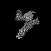
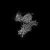
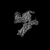
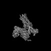
| 登録情報 | データベース: PDB / ID: 7y64 | ||||||
|---|---|---|---|---|---|---|---|
| タイトル | Cryo-EM structure of C5a-bound C5aR1 in complex with Gi protein | ||||||
 要素 要素 |
| ||||||
 キーワード キーワード | MEMBRANE PROTEIN / Complex / Agonist | ||||||
| 機能・相同性 |  機能・相同性情報 機能・相同性情報presynapse organization / complement component C5a signaling pathway / Terminal pathway of complement / complement component C5a receptor activity / membrane attack complex / response to peptidoglycan / Activation of C3 and C5 / complement activation, GZMK pathway / negative regulation of macrophage chemotaxis / sensory perception of chemical stimulus ...presynapse organization / complement component C5a signaling pathway / Terminal pathway of complement / complement component C5a receptor activity / membrane attack complex / response to peptidoglycan / Activation of C3 and C5 / complement activation, GZMK pathway / negative regulation of macrophage chemotaxis / sensory perception of chemical stimulus / other organism cell membrane / complement receptor mediated signaling pathway / complement activation, alternative pathway / positive regulation of neutrophil chemotaxis / chemokine activity / endopeptidase inhibitor activity / positive regulation of macrophage chemotaxis / amyloid-beta clearance / complement activation, classical pathway / positive regulation of vascular endothelial growth factor production / cellular defense response / positive regulation of chemokine production / neutrophil chemotaxis / adenylate cyclase inhibitor activity / positive regulation of protein localization to cell cortex / T cell migration / positive regulation of relaxation of smooth muscle / Adenylate cyclase inhibitory pathway / D2 dopamine receptor binding / adenylate cyclase-inhibiting serotonin receptor signaling pathway / G protein-coupled serotonin receptor binding / astrocyte activation / Regulation of Complement cascade / secretory granule membrane / cellular response to forskolin / positive regulation of epithelial cell proliferation / Peptide ligand-binding receptors / regulation of mitotic spindle organization / chemokine-mediated signaling pathway / Regulation of insulin secretion / neuropeptide signaling pathway / response to prostaglandin E / microglial cell activation / positive regulation of cholesterol biosynthetic process / negative regulation of insulin secretion / mRNA transcription by RNA polymerase II / G protein-coupled receptor binding / response to peptide hormone / cognition / G protein-coupled receptor activity / chemotaxis / centriolar satellite / G-protein beta/gamma-subunit complex binding / positive regulation of angiogenesis / adenylate cyclase-modulating G protein-coupled receptor signaling pathway / apical part of cell / adenylate cyclase-inhibiting G protein-coupled receptor signaling pathway / Olfactory Signaling Pathway / Activation of the phototransduction cascade / G protein-coupled acetylcholine receptor signaling pathway / G beta:gamma signalling through PLC beta / Presynaptic function of Kainate receptors / Thromboxane signalling through TP receptor / Activation of G protein gated Potassium channels / Inhibition of voltage gated Ca2+ channels via Gbeta/gamma subunits / G-protein activation / Glucagon signaling in metabolic regulation / Prostacyclin signalling through prostacyclin receptor / G beta:gamma signalling through CDC42 / Synthesis, secretion, and inactivation of Glucagon-like Peptide-1 (GLP-1) / G beta:gamma signalling through BTK / photoreceptor disc membrane / ADP signalling through P2Y purinoceptor 12 / Sensory perception of sweet, bitter, and umami (glutamate) taste / Glucagon-type ligand receptors / GDP binding / Adrenaline,noradrenaline inhibits insulin secretion / Vasopressin regulates renal water homeostasis via Aquaporins / Glucagon-like Peptide-1 (GLP1) regulates insulin secretion / G alpha (z) signalling events / cellular response to catecholamine stimulus / ADP signalling through P2Y purinoceptor 1 / ADORA2B mediated anti-inflammatory cytokines production / G beta:gamma signalling through PI3Kgamma / adenylate cyclase-activating dopamine receptor signaling pathway / Cooperation of PDCL (PhLP1) and TRiC/CCT in G-protein beta folding / GPER1 signaling / cellular response to prostaglandin E stimulus / heterotrimeric G-protein complex / G alpha (12/13) signalling events / Inactivation, recovery and regulation of the phototransduction cascade / G-protein beta-subunit binding / extracellular vesicle / sensory perception of taste / sperm principal piece / Thrombin signalling through proteinase activated receptors (PARs) / signaling receptor complex adaptor activity / adenylate cyclase-activating G protein-coupled receptor signaling pathway / positive regulation of cytosolic calcium ion concentration / retina development in camera-type eye 類似検索 - 分子機能 | ||||||
| 生物種 |  Homo sapiens (ヒト) Homo sapiens (ヒト) | ||||||
| 手法 | 電子顕微鏡法 / 単粒子再構成法 / クライオ電子顕微鏡法 / 解像度: 2.9 Å | ||||||
 データ登録者 データ登録者 | Feng, Y.Y. / Zhao, C. / Yan, W. / Shao, Z.H. | ||||||
| 資金援助 |  中国, 1件 中国, 1件
| ||||||
 引用 引用 | ジャーナル: Cell Res / 年: 2023 タイトル: Mechanism of activation and biased signaling in complement receptor C5aR1. 著者: Yuying Feng / Chang Zhao / Yue Deng / Heli Wang / Liang Ma / Sicen Liu / Xiaowen Tian / Bo Wang / Yan Bin / Peipei Chen / Wei Yan / Ping Fu / Zhenhua Shao / 要旨: The complement system plays an important role in the innate immune response to invading pathogens. The complement fragment C5a is one of its important effector components and exerts diverse ...The complement system plays an important role in the innate immune response to invading pathogens. The complement fragment C5a is one of its important effector components and exerts diverse physiological functions through activation of the C5a receptor 1 (C5aR1) and associated downstream G protein and β-arrestin signaling pathways. Dysfunction of the C5a-C5aR1 axis is linked to numerous inflammatory and immune-mediated diseases, but the structural basis for activation and biased signaling of C5aR1 remains elusive. Here, we present cryo-electron microscopy structures of the activated wild-type C5aR1-G protein complex bound to each of the following: C5a, the hexapeptidic agonist C5a, and the G protein-biased agonist BM213. The structures reveal the landscape of the C5a-C5aR1 interaction as well as a common motif for the recognition of diverse orthosteric ligands. Moreover, combined with mutagenesis studies and cell-based pharmacological assays, we deciphered a framework for biased signaling using different peptide analogs and provided insight into the activation mechanism of C5aR1 by solving the structure of C5aR1 mutant-G signaling activation complex induced by C089, which exerts antagonism on wild-type C5aR1. In addition, unusual conformational changes in the intracellular end of transmembrane domain 7 and helix 8 upon agonist binding suggest a differential signal transduction process. Collectively, our study provides mechanistic understanding into the ligand recognition, biased signaling modulation, activation, and G protein coupling of C5aR1, which may facilitate the future design of therapeutic agents. | ||||||
| 履歴 |
|
- 構造の表示
構造の表示
| 構造ビューア | 分子: MolmilJmol/JSmol |
|---|
- ダウンロードとリンク
-ダウンロード
| PDBx/mmCIF形式 | 7y64.cif.gz | 243.9 KB | 表示 | PDBx/mmCIF形式 |
|---|---|---|---|---|
| PDB形式 | pdb7y64.ent.gz | 187.4 KB | 表示 | PDB形式 |
| PDBx/mmJSON形式 | 7y64.json.gz | ツリー表示 | PDBx/mmJSON形式 | |
| その他 |  その他のダウンロード その他のダウンロード |
-検証レポート
| アーカイブディレクトリ | https://data.pdbj.org/pub/pdb/validation_reports/y6/7y64ftp://data.pdbj.org/pub/pdb/validation_reports/y6/7y64 | HTTPS FTP |
|---|
-関連構造データ


-リンク
 PDBj
PDBj


- 集合体
集合体
| 登録構造単位 | 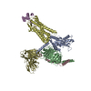
|
|---|---|
| 1 |
|
-要素
-Guanine nucleotide-binding protein ... , 3種, 3分子 ABC
| #1: タンパク質 | 分子量: 40415.031 Da / 分子数: 1 / 由来タイプ: 組換発現 / 由来: (組換発現) Homo sapiens (ヒト) / 遺伝子: GNAI1発現宿主:   Spodoptera frugiperda (ツマジロクサヨトウ) Spodoptera frugiperda (ツマジロクサヨトウ)参照: UniProt: P63096 |
|---|---|
| #2: タンパク質 | 分子量: 39141.793 Da / 分子数: 1 / 由来タイプ: 組換発現 / 由来: (組換発現) Homo sapiens (ヒト) / 遺伝子: GNB1発現宿主: Spodoptera frugiperda (ツマジロクサヨトウ)参照: UniProt: P62873 |
| #3: タンパク質 | 分子量: 7861.143 Da / 分子数: 1 / 由来タイプ: 組換発現 / 由来: (組換発現) Homo sapiens (ヒト) / 遺伝子: GNG2発現宿主: Spodoptera frugiperda (ツマジロクサヨトウ)参照: UniProt: P59768 |
-タンパク質 , 2種, 2分子 DE
| #5: タンパク質 | 分子量: 38415.238 Da / 分子数: 1 / 由来タイプ: 組換発現 / 由来: (組換発現) Homo sapiens (ヒト) / 遺伝子: C5AR1, C5AR, C5R1発現宿主: Spodoptera frugiperda (ツマジロクサヨトウ)参照: UniProt: P21730 |
|---|---|
| #6: タンパク質 | 分子量: 8342.727 Da / 分子数: 1 / 由来タイプ: 組換発現 / 由来: (組換発現) Homo sapiens (ヒト) / 遺伝子: C5, CPAMD4 / 発現宿主:  |
-抗体 , 1種, 1分子 S
| #4: 抗体 | 分子量: 28636.793 Da / 分子数: 1 / 由来タイプ: 組換発現 / 由来: (組換発現) 発現宿主: Spodoptera frugiperda (ツマジロクサヨトウ) |
|---|
-詳細
| Has protein modification | Y |
|---|
-実験情報
-実験
| 実験 | 手法: 電子顕微鏡法 |
|---|---|
| EM実験 | 試料の集合状態: PARTICLE / 3次元再構成法: 単粒子再構成法 |
- 試料調製
試料調製
| 構成要素 |
| ||||||||||||||||||||||||||||||
|---|---|---|---|---|---|---|---|---|---|---|---|---|---|---|---|---|---|---|---|---|---|---|---|---|---|---|---|---|---|---|---|
| 由来(天然) |
| ||||||||||||||||||||||||||||||
| 由来(組換発現) |
| ||||||||||||||||||||||||||||||
| 緩衝液 | pH: 7.5 | ||||||||||||||||||||||||||||||
| 試料 | 包埋: NO / シャドウイング: NO / 染色: NO / 凍結: YES | ||||||||||||||||||||||||||||||
| 急速凍結 | 凍結剤: ETHANE |
- 電子顕微鏡撮影
| 実験機器 |  モデル: Titan Krios / 画像提供: FEI Company |
|---|---|
| 顕微鏡 | モデル: FEI TITAN KRIOS |
| 電子銃 | 電子線源:  FIELD EMISSION GUN / 加速電圧: 300 kV / 照射モード: FLOOD BEAM FIELD EMISSION GUN / 加速電圧: 300 kV / 照射モード: FLOOD BEAM |
| 電子レンズ | モード: BRIGHT FIELD / 最大 デフォーカス(公称値): 1800 nm / 最小 デフォーカス(公称値): 1000 nm / Cs: 2.7 mm |
| 撮影 | 電子線照射量: 66 e/Å2 フィルム・検出器のモデル: GATAN K2 SUMMIT (4k x 4k) |
- 解析
解析
| CTF補正 | タイプ: PHASE FLIPPING AND AMPLITUDE CORRECTION |
|---|---|
| 3次元再構成 | 解像度: 2.9 Å / 解像度の算出法: FSC 0.143 CUT-OFF / 粒子像の数: 623262 / 対称性のタイプ: POINT |
