 Movie
Movie Controller

[English] 日本語
 Yorodumi
- PDB-7y1l: Structure of SUR2B in complex with Mg-ATP and repaglinide in the ... -
+ Open data

- Basic information
| Entry | Database: PDB / ID: 7y1l | ||||||||||||
|---|---|---|---|---|---|---|---|---|---|---|---|---|---|
| Title | Structure of SUR2B in complex with Mg-ATP and repaglinide in the inward-facing conformation | ||||||||||||
 Components Components | Isoform SUR2B of ATP-binding cassette sub-family C member 9 | ||||||||||||
 Keywords Keywords | MEMBRANE PROTEIN / SUR2B / ABC transporter / repaglinide | ||||||||||||
| Function / homology |  Function and homology information Function and homology informationvascular process in circulatory system / substrate-dependent cell migration, cell contraction / oxygen metabolic process / reactive oxygen species biosynthetic process / ATP sensitive Potassium channels / response to decreased oxygen levels / ABC-family protein mediated transport / potassium channel activator activity / inward rectifying potassium channel / sulfonylurea receptor activity ...vascular process in circulatory system / substrate-dependent cell migration, cell contraction / oxygen metabolic process / reactive oxygen species biosynthetic process / ATP sensitive Potassium channels / response to decreased oxygen levels / ABC-family protein mediated transport / potassium channel activator activity / inward rectifying potassium channel / sulfonylurea receptor activity / response to potassium ion / response to peptide / cardiac conduction / response to oxygen levels / ATPase-coupled monoatomic cation transmembrane transporter activity / response to hydrogen sulfide / coronary vasculature development / cellular response to chemical stress / regulation of potassium ion transmembrane transport / cellular response to potassium ion / circulatory system development / cellular respiration / syntaxin binding / heterocyclic compound binding / : / Ion homeostasis / cardiac muscle cell contraction / blood circulation / blood vessel development / fatty acid oxidation / response to ATP / cellular response to ATP / potassium ion import across plasma membrane / response to stress / ATPase-coupled transmembrane transporter activity / monoatomic cation transmembrane transport / action potential / potassium channel regulator activity / potassium channel activity / ABC-type transporter activity / skeletal muscle tissue development / ATP metabolic process / heart morphogenesis / negative regulation of blood pressure / T-tubule / potassium ion transmembrane transport / cellular response to calcium ion / acrosomal vesicle / blood vessel diameter maintenance / response to activity / sarcomere / response to hydrogen peroxide / potassium ion transport / mitochondrion organization / regulation of membrane potential / sarcolemma / response to estrogen / regulation of blood pressure / cellular response to xenobiotic stimulus / vasodilation / MAPK cascade / heart development / fibroblast proliferation / gene expression / defense response to virus / transmembrane transporter binding / response to hypoxia / response to xenobiotic stimulus / regulation of transcription by RNA polymerase II / negative regulation of apoptotic process / protein-containing complex binding / ATP hydrolysis activity / protein-containing complex / mitochondrion / ATP binding / identical protein binding / plasma membrane / cytoplasm Similarity search - Function | ||||||||||||
| Biological species |  | ||||||||||||
| Method | ELECTRON MICROSCOPY / single particle reconstruction / cryo EM / Resolution: 3.73 Å | ||||||||||||
 Authors Authors | Chen, L. / Ding, D. / Hou, T. | ||||||||||||
| Funding support |  China, 3items China, 3items
| ||||||||||||
 Citation Citation | Journal: Nat Commun / Year: 2023 Title: The inhibition mechanism of the SUR2A-containing K channel by a regulatory helix. Authors: Dian Ding / Tianyi Hou / Miao Wei / Jing-Xiang Wu / Lei Chen / Abstract: K channels are metabolic sensors for intracellular ATP/ADP ratios, play essential roles in many physiological processes, and are implicated in a spectrum of pathological conditions. SUR2A-containing ...K channels are metabolic sensors for intracellular ATP/ADP ratios, play essential roles in many physiological processes, and are implicated in a spectrum of pathological conditions. SUR2A-containing K channels differ from other subtypes in their sensitivity to Mg-ADP activation. However, the underlying structural mechanism remains poorly understood. Here we present a series of cryo-EM structures of SUR2A in the presence of different combinations of Mg-nucleotides and the allosteric inhibitor repaglinide. These structures uncover regulatory helix (R helix) on the NBD1-TMD2 linker, which wedges between NBD1 and NBD2. R helix stabilizes SUR2A in the NBD-separated conformation to inhibit channel activation. The competitive binding of Mg-ADP with Mg-ATP to NBD2 mobilizes the R helix to relieve such inhibition, allowing channel activation. The structures of SUR2B in similar conditions suggest that the C-terminal 42 residues of SUR2B enhance the structural dynamics of NBD2 and facilitate the dissociation of the R helix and the binding of Mg-ADP to NBD2, promoting NBD dimerization and subsequent channel activation. | ||||||||||||
| History |
|
- Structure visualization
| Structure viewer | Molecule: MolmilJmol/JSmol |
|---|
- Downloads & links
-Download
| PDBx/mmCIF format | 7y1l.cif.gz | 221.3 KB | Display | PDBx/mmCIF format |
|---|---|---|---|---|
| PDB format | pdb7y1l.ent.gz | 167.5 KB | Display | PDB format |
| PDBx/mmJSON format | 7y1l.json.gz | Tree view | PDBx/mmJSON format | |
| Others |  Other downloads Other downloads |
-Validation report
| Arichive directory | https://data.pdbj.org/pub/pdb/validation_reports/y1/7y1lftp://data.pdbj.org/pub/pdb/validation_reports/y1/7y1l | HTTPS FTP |
|---|
-Related structure data
| Related structure data |  33565MC 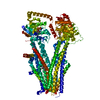 7y1jC 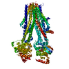 7y1kC 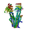 7y1mC 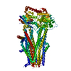 7y1nC M: map data used to model this data C: citing same article ( |
|---|---|
| Similar structure data |
-Links
 PDBj
PDBj


- Assembly
| Deposited unit | 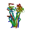
|
|---|---|
| 1 |
|
-Components
| #1: Protein | Mass: 174488.562 Da / Num. of mol.: 1 Source method: isolated from a genetically manipulated source Source: (gene. exp.)  Homo sapiens (human) / References: UniProt: Q63563 Homo sapiens (human) / References: UniProt: Q63563 | ||||||
|---|---|---|---|---|---|---|---|
| #2: Chemical |   Mass: 24.305 Da / Num. of mol.: 2 / Source method: obtained synthetically / Formula: Mg / Feature type: SUBJECT OF INVESTIGATION Mass: 24.305 Da / Num. of mol.: 2 / Source method: obtained synthetically / Formula: Mg / Feature type: SUBJECT OF INVESTIGATION#3: Chemical |   Mass: 507.181 Da / Num. of mol.: 2 / Source method: obtained synthetically / Formula: C10H16N5O13P3 / Feature type: SUBJECT OF INVESTIGATION / Comment: ATP, energy-carrying molecule*YM Mass: 507.181 Da / Num. of mol.: 2 / Source method: obtained synthetically / Formula: C10H16N5O13P3 / Feature type: SUBJECT OF INVESTIGATION / Comment: ATP, energy-carrying molecule*YM#4: Chemical | ChemComp-BJX / |   Mass: 452.586 Da / Num. of mol.: 1 / Source method: obtained synthetically / Formula: C27H36N2O4 Mass: 452.586 Da / Num. of mol.: 1 / Source method: obtained synthetically / Formula: C27H36N2O4Has ligand of interest | Y | |
-Experimental details
-Experiment
| Experiment | Method: ELECTRON MICROSCOPY |
|---|---|
| EM experiment | Aggregation state: PARTICLE / 3D reconstruction method: single particle reconstruction |
- Sample preparation
| Component | Name: ATP-binding cassette sub-family C member 9, isoform B / Type: COMPLEX / Entity ID: #1 / Source: RECOMBINANT |
|---|---|
| Molecular weight | Value: 174 kDa/nm / Experimental value: NO |
| Source (natural) | Organism: |
| Source (recombinant) | Organism: Homo sapiens (human) |
| Buffer solution | pH: 7.5 |
| Specimen | Embedding applied: NO / Shadowing applied: NO / Staining applied: NO / Vitrification applied: YES |
| Vitrification | Cryogen name: ETHANE |
- Electron microscopy imaging
| Experimental equipment |  Model: Titan Krios / Image courtesy: FEI Company |
|---|---|
| Microscopy | Model: FEI TITAN KRIOS |
| Electron gun | Electron source:  FIELD EMISSION GUN / Accelerating voltage: 300 kV / Illumination mode: FLOOD BEAM FIELD EMISSION GUN / Accelerating voltage: 300 kV / Illumination mode: FLOOD BEAM |
| Electron lens | Mode: BRIGHT FIELD / Nominal defocus max: 2000 nm / Nominal defocus min: 1800 nm |
| Image recording | Electron dose: 52 e/Å2 / Film or detector model: GATAN K3 (6k x 4k) |
- Processing
| EM software | Name: cryoSPARC / Category: 3D reconstruction |
|---|---|
| CTF correction | Type: PHASE FLIPPING AND AMPLITUDE CORRECTION |
| 3D reconstruction | Resolution: 3.73 Å / Resolution method: FSC 0.143 CUT-OFF / Num. of particles: 60550 / Symmetry type: POINT |
