 Movie
Movie Controller

[English] 日本語
 Yorodumi
- PDB-7xxh: Cryo-EM structure of the purinergic receptor P2Y1R in complex wit... -
+ Open data

- Basic information
| Entry | Database: PDB / ID: 7xxh | |||||||||||||||||||||||||||||||||||||||||||||
|---|---|---|---|---|---|---|---|---|---|---|---|---|---|---|---|---|---|---|---|---|---|---|---|---|---|---|---|---|---|---|---|---|---|---|---|---|---|---|---|---|---|---|---|---|---|---|
| Title | Cryo-EM structure of the purinergic receptor P2Y1R in complex with 2MeSADP and G11 | |||||||||||||||||||||||||||||||||||||||||||||
 Components Components |
| |||||||||||||||||||||||||||||||||||||||||||||
 Keywords Keywords | MEMBRANE PROTEIN / G protein-coupled receptor / purinergic receptor / P2Y1R / ligand binding / signal transduction | |||||||||||||||||||||||||||||||||||||||||||||
| Function / homology |  Function and homology information Function and homology informationG protein-coupled ATP receptor activity / cellular response to purine-containing compound / relaxation of muscle / G protein-coupled ADP receptor activity / A1 adenosine receptor binding / G protein-coupled purinergic nucleotide receptor signaling pathway / positive regulation of inositol trisphosphate biosynthetic process / P2Y receptors / positive regulation of penile erection / G protein-coupled purinergic nucleotide receptor activity ...G protein-coupled ATP receptor activity / cellular response to purine-containing compound / relaxation of muscle / G protein-coupled ADP receptor activity / A1 adenosine receptor binding / G protein-coupled purinergic nucleotide receptor signaling pathway / positive regulation of inositol trisphosphate biosynthetic process / P2Y receptors / positive regulation of penile erection / G protein-coupled purinergic nucleotide receptor activity / negative regulation of norepinephrine secretion / positive regulation of monoatomic ion transport / glial cell migration / signaling receptor regulator activity / regulation of presynaptic cytosolic calcium ion concentration / G protein-coupled adenosine receptor signaling pathway / response to growth factor / positive regulation of hormone secretion / signal transduction involved in regulation of gene expression / eating behavior / regulation of synaptic vesicle exocytosis / cellular response to ATP / response to mechanical stimulus / monoatomic ion transport / presynaptic active zone membrane / blood vessel diameter maintenance / protein localization to plasma membrane / establishment of localization in cell / ADP binding / platelet activation / adenylate cyclase-inhibiting G protein-coupled receptor signaling pathway / Olfactory Signaling Pathway / Activation of the phototransduction cascade / G beta:gamma signalling through PLC beta / Presynaptic function of Kainate receptors / Thromboxane signalling through TP receptor / G protein-coupled acetylcholine receptor signaling pathway / Activation of G protein gated Potassium channels / Inhibition of voltage gated Ca2+ channels via Gbeta/gamma subunits / G-protein activation / G beta:gamma signalling through CDC42 / Prostacyclin signalling through prostacyclin receptor / Glucagon signaling in metabolic regulation / G beta:gamma signalling through BTK / Synthesis, secretion, and inactivation of Glucagon-like Peptide-1 (GLP-1) / ADP signalling through P2Y purinoceptor 12 / photoreceptor disc membrane / Sensory perception of sweet, bitter, and umami (glutamate) taste / Glucagon-type ligand receptors / Adrenaline,noradrenaline inhibits insulin secretion / Vasopressin regulates renal water homeostasis via Aquaporins / Glucagon-like Peptide-1 (GLP1) regulates insulin secretion / G alpha (z) signalling events / ADP signalling through P2Y purinoceptor 1 / cellular response to catecholamine stimulus / ADORA2B mediated anti-inflammatory cytokines production / G beta:gamma signalling through PI3Kgamma / adenylate cyclase-activating dopamine receptor signaling pathway / Cooperation of PDCL (PhLP1) and TRiC/CCT in G-protein beta folding / GPER1 signaling / regulation of cell shape / signaling receptor activity / G-protein beta-subunit binding / cellular response to prostaglandin E stimulus / heterotrimeric G-protein complex / Inactivation, recovery and regulation of the phototransduction cascade / G alpha (12/13) signalling events / extracellular vesicle / sensory perception of taste / Thrombin signalling through proteinase activated receptors (PARs) / signaling receptor complex adaptor activity / positive regulation of cytosolic calcium ion concentration / retina development in camera-type eye / cell body / GTPase binding / Ca2+ pathway / scaffold protein binding / fibroblast proliferation / High laminar flow shear stress activates signaling by PIEZO1 and PECAM1:CDH5:KDR in endothelial cells / G alpha (i) signalling events / basolateral plasma membrane / G alpha (s) signalling events / phospholipase C-activating G protein-coupled receptor signaling pathway / G alpha (q) signalling events / postsynaptic membrane / Ras protein signal transduction / cell surface receptor signaling pathway / Extra-nuclear estrogen signaling / positive regulation of ERK1 and ERK2 cascade / cell population proliferation / postsynaptic density / cilium / apical plasma membrane / G protein-coupled receptor signaling pathway / protein heterodimerization activity / lysosomal membrane / GTPase activity / dendrite / synapse / protein-containing complex binding Similarity search - Function | |||||||||||||||||||||||||||||||||||||||||||||
| Biological species |  Homo sapiens (human) Homo sapiens (human) | |||||||||||||||||||||||||||||||||||||||||||||
| Method | ELECTRON MICROSCOPY / single particle reconstruction / cryo EM / Resolution: 2.9 Å | |||||||||||||||||||||||||||||||||||||||||||||
 Authors Authors | Tan, Q. / Li, B. / Han, S. / Zhao, Q. / Wu, B. | |||||||||||||||||||||||||||||||||||||||||||||
| Funding support |  China, 5items China, 5items
| |||||||||||||||||||||||||||||||||||||||||||||
 Citation Citation | Journal: Protein Cell / Year: 2023 Title: Structural insights into signal transduction of the purinergic receptors P2Y1R and P2Y12R. Authors: Beibei Li / Shuo Han / Mu Wang / Yu Yu / Limin Ma / Xiaojing Chu / Qiuxiang Tan / Qiang Zhao / Beili Wu / | |||||||||||||||||||||||||||||||||||||||||||||
| History |
|
- Structure visualization
| Structure viewer | Molecule: MolmilJmol/JSmol |
|---|
- Downloads & links
-Download
| PDBx/mmCIF format | 7xxh.cif.gz | 222 KB | Display | PDBx/mmCIF format |
|---|---|---|---|---|
| PDB format | pdb7xxh.ent.gz | 164.9 KB | Display | PDB format |
| PDBx/mmJSON format | 7xxh.json.gz | Tree view | PDBx/mmJSON format | |
| Others |  Other downloads Other downloads |
-Validation report
| Arichive directory | https://data.pdbj.org/pub/pdb/validation_reports/xx/7xxhftp://data.pdbj.org/pub/pdb/validation_reports/xx/7xxh | HTTPS FTP |
|---|
-Related structure data
| Related structure data |  33503MC 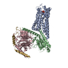 7xxiC M: map data used to model this data C: citing same article ( |
|---|---|
| Similar structure data |

-Links
 PDBj
PDBj


- Assembly
| Deposited unit | 
|
|---|---|
| 1 |
|
-Components
-Guanine nucleotide-binding protein ... , 3 types, 3 molecules ABG
| #2: Protein | Mass: 41385.281 Da / Num. of mol.: 1 Source method: isolated from a genetically manipulated source Source: (gene. exp.) Homo sapiens (human) / Production host:   Spodoptera frugiperda (fall armyworm) Spodoptera frugiperda (fall armyworm) |
|---|---|
| #3: Protein | Mass: 38245.805 Da / Num. of mol.: 1 Source method: isolated from a genetically manipulated source Source: (gene. exp.) Homo sapiens (human) / Gene: GNB1 / Production host: Spodoptera frugiperda (fall armyworm) / References: UniProt: P62873 |
| #4: Protein | Mass: 7861.143 Da / Num. of mol.: 1 Source method: isolated from a genetically manipulated source Source: (gene. exp.) Homo sapiens (human) / Gene: GNG2 / Production host: Spodoptera frugiperda (fall armyworm) / References: UniProt: P59768 |
-Protein / Antibody / Non-polymers , 3 types, 3 molecules RH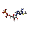
| #1: Protein | Mass: 46296.887 Da / Num. of mol.: 1 Source method: isolated from a genetically manipulated source Source: (gene. exp.) Homo sapiens (human) / Gene: P2RY1 / Production host: Spodoptera frugiperda (fall armyworm) / References: UniProt: P47900 |
|---|---|
| #5: Antibody | Mass: 26337.307 Da / Num. of mol.: 1 Source method: isolated from a genetically manipulated source Source: (gene. exp.) Homo sapiens (human) / Production host: Spodoptera frugiperda (fall armyworm) |
| #6: Chemical | ChemComp-6AD /  Mass: 473.293 Da / Num. of mol.: 1 / Source method: obtained synthetically / Formula: C11H17N5O10P2S / Feature type: SUBJECT OF INVESTIGATION Mass: 473.293 Da / Num. of mol.: 1 / Source method: obtained synthetically / Formula: C11H17N5O10P2S / Feature type: SUBJECT OF INVESTIGATION |
-Details
| Has ligand of interest | Y |
|---|---|
| Has protein modification | Y |
-Experimental details
-Experiment
| Experiment | Method: ELECTRON MICROSCOPY |
|---|---|
| EM experiment | Aggregation state: PARTICLE / 3D reconstruction method: single particle reconstruction |
- Sample preparation
| Component | Name: The purinergic receptor P2Y1R in complex with 2MeSADP, G11 and scfv16 Type: COMPLEX / Details: scf16 was recombinantly expressed in sf9 cells / Entity ID: #2-#5 / Source: RECOMBINANT |
|---|---|
| Molecular weight | Value: 10 kDa/nm / Experimental value: NO |
| Source (natural) | Organism: Homo sapiens (human) |
| Source (recombinant) | Organism: Spodoptera frugiperda (fall armyworm) |
| Buffer solution | pH: 7.5 |
| Buffer component | Conc.: 2.0 mg/ml / Name: sodium chloride / Formula: 150mM |
| Specimen | Conc.: 2 mg/ml / Embedding applied: NO / Shadowing applied: NO / Staining applied: NO / Vitrification applied: YES / Details: This sample was monodisperse |
| Specimen support | Grid material: GOLD / Grid mesh size: 400 divisions/in. / Grid type: C-flat-1.2/1.3 |
| Vitrification | Instrument: FEI VITROBOT MARK IV / Cryogen name: ETHANE / Humidity: 100 % / Chamber temperature: 298 K / Details: blot for 1s |
- Electron microscopy imaging
| Experimental equipment |  Model: Titan Krios / Image courtesy: FEI Company |
|---|---|
| Microscopy | Model: FEI TITAN KRIOS |
| Electron gun | Electron source:  FIELD EMISSION GUN / Accelerating voltage: 300 kV / Illumination mode: FLOOD BEAM FIELD EMISSION GUN / Accelerating voltage: 300 kV / Illumination mode: FLOOD BEAM |
| Electron lens | Mode: BRIGHT FIELD / Nominal defocus max: 1500 nm / Nominal defocus min: 800 nm / Cs: 2.7 mm / Alignment procedure: BASIC |
| Image recording | Average exposure time: 2 sec. / Electron dose: 60 e/Å2 / Film or detector model: GATAN K3 BIOQUANTUM (6k x 4k) / Num. of grids imaged: 2 / Num. of real images: 9856 / Details: Images were collected in movie-mode |
- Processing
| EM software |
| ||||||||||||||||||||||||
|---|---|---|---|---|---|---|---|---|---|---|---|---|---|---|---|---|---|---|---|---|---|---|---|---|---|
| Image processing | Details: The selected images were 20eV using GIF-Quantum LS Imaging energy filter | ||||||||||||||||||||||||
| CTF correction | Type: NONE | ||||||||||||||||||||||||
| Particle selection | Num. of particles selected: 6184568 | ||||||||||||||||||||||||
| Symmetry | Point symmetry: C1 (asymmetric) | ||||||||||||||||||||||||
| 3D reconstruction | Resolution: 2.9 Å / Resolution method: FSC 0.143 CUT-OFF / Num. of particles: 659399 / Symmetry type: POINT | ||||||||||||||||||||||||
| Atomic model building | B value: 43.86 / Space: REAL | ||||||||||||||||||||||||
| Atomic model building | PDB-ID: 4NXW Accession code: 4NXW / Source name: PDB / Type: experimental model | ||||||||||||||||||||||||
| Refinement | Stereochemistry target values: GeoStd + Monomer Library + CDL v1.2 | ||||||||||||||||||||||||
| Displacement parameters | Biso mean: 43.9 Å2 | ||||||||||||||||||||||||
| Refine LS restraints |
|

