[English] 日本語
 Yorodumi
- PDB-7xg4: CryoEM structure of type IV-A CasDinG bound NTS-nicked Csf-crRNA-... -
+ Open data
- Basic information
| Entry | Database: PDB / ID: 7xg4 | ||||||
|---|---|---|---|---|---|---|---|
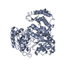
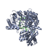
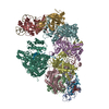
| Title | CryoEM structure of type IV-A CasDinG bound NTS-nicked Csf-crRNA-dsDNA quaternary complex in a second state | ||||||
 Components Components |
| ||||||
 Keywords Keywords | STRUCTURAL PROTEIN/RNA/DNA / Nuclease / STRUCTURAL PROTEIN-RNA-DNA COMPLEX | ||||||
| Function / homology | DNA / DNA (> 10) / RNA / RNA (> 10) Function and homology information Function and homology information | ||||||
| Biological species |  | ||||||
| Method | ELECTRON MICROSCOPY / single particle reconstruction / cryo EM / Resolution: 3.7 Å | ||||||
 Authors Authors | Zhang, J.T. / Cui, N. / Huang, H.D. / Jia, N. | ||||||
| Funding support | 1items
| ||||||
 Citation Citation |  Journal: Mol Cell / Year: 2023 Journal: Mol Cell / Year: 2023Title: Type IV-A CRISPR-Csf complex: Assembly, dsDNA targeting, and CasDinG recruitment. Authors: Ning Cui / Jun-Tao Zhang / Yongrui Liu / Yanhong Liu / Xiao-Yu Liu / Chongyuan Wang / Hongda Huang / Ning Jia /  Abstract: Type IV CRISPR-Cas systems, which are primarily found on plasmids and exhibit a strong plasmid-targeting preference, are the only one of the six known CRISPR-Cas types for which the mechanistic ...Type IV CRISPR-Cas systems, which are primarily found on plasmids and exhibit a strong plasmid-targeting preference, are the only one of the six known CRISPR-Cas types for which the mechanistic details of their function remain unknown. Here, we provide high-resolution functional snapshots of type IV-A Csf complexes before and after target dsDNA binding, either in the absence or presence of CasDinG, revealing the mechanisms underlying Csf complex assembly, "DWN" PAM-dependent dsDNA targeting, R-loop formation, and CasDinG recruitment. Furthermore, we establish that CasDinG, a signature DinG family helicase, harbors ssDNA-stimulated ATPase activity and ATP-dependent 5'-3' DNA helicase activity. In addition, we show that CasDinG unwinds the non-target strand (NTS) and target strand (TS) of target dsDNA from the Csf complex. These molecular details advance our mechanistic understanding of type IV-A CRISPR-Csf function and should enable Csf complexes to be harnessed as genome-engineering tools for biotechnological applications. | ||||||
| History |
|
- Structure visualization
| Structure viewer | Molecule:  Molmil Molmil Jmol/JSmol Jmol/JSmol |
|---|
- Downloads & links
- Download
| PDBx/mmCIF format |  7xg4.cif.gz 7xg4.cif.gz | 626 KB | Display |  PDBx/mmCIF format PDBx/mmCIF format |
|---|---|---|---|---|
| PDB format |  pdb7xg4.ent.gz pdb7xg4.ent.gz | 472.3 KB | Display |  PDB format PDB format |
| PDBx/mmJSON format |  7xg4.json.gz 7xg4.json.gz | Tree view |  PDBx/mmJSON format PDBx/mmJSON format | |
| Others |  Other downloads Other downloads |
-Validation report
| Summary document |  7xg4_validation.pdf.gz 7xg4_validation.pdf.gz | 1.4 MB | Display |  wwPDB validaton report wwPDB validaton report |
|---|---|---|---|---|
| Full document |  7xg4_full_validation.pdf.gz 7xg4_full_validation.pdf.gz | 1.5 MB | Display | |
| Data in XML |  7xg4_validation.xml.gz 7xg4_validation.xml.gz | 108.2 KB | Display | |
| Data in CIF |  7xg4_validation.cif.gz 7xg4_validation.cif.gz | 159.5 KB | Display | |
| Arichive directory |  https://data.pdbj.org/pub/pdb/validation_reports/xg/7xg4 https://data.pdbj.org/pub/pdb/validation_reports/xg/7xg4 ftp://data.pdbj.org/pub/pdb/validation_reports/xg/7xg4 ftp://data.pdbj.org/pub/pdb/validation_reports/xg/7xg4 | HTTPS FTP |
-Related structure data
| Related structure data |  33185MC  7xexC  7xf0C  7xf1C  7xfzC  7xg0C  7xg2C  7xg3C M: map data used to model this data C: citing same article ( |
|---|---|
| Similar structure data | Similarity search - Function & homology  F&H Search F&H Search |
- Links
Links
- Assembly
| Deposited unit | 
|
|---|---|
| 1 |
|
- Components
-Protein , 5 types, 9 molecules ABCDEFGHL
| #1: Protein | Mass: 28675.768 Da / Num. of mol.: 1 Source method: isolated from a genetically manipulated source Source: (gene. exp.)   | ||||
|---|---|---|---|---|---|
| #2: Protein | Mass: 24421.953 Da / Num. of mol.: 1 Source method: isolated from a genetically manipulated source Source: (gene. exp.)   | ||||
| #3: Protein | Mass: 37322.234 Da / Num. of mol.: 5 Source method: isolated from a genetically manipulated source Source: (gene. exp.)   #4: Protein | | Mass: 29152.414 Da / Num. of mol.: 1 Source method: isolated from a genetically manipulated source Source: (gene. exp.)   #8: Protein | | Mass: 67939.883 Da / Num. of mol.: 1 Source method: isolated from a genetically manipulated source Source: (gene. exp.)   |
-DNA chain , 2 types, 2 molecules JK
| #6: DNA chain | Mass: 11520.440 Da / Num. of mol.: 1 / Source method: obtained synthetically / Source: (synth.)  |
|---|---|
| #7: DNA chain | Mass: 16547.568 Da / Num. of mol.: 1 / Source method: obtained synthetically / Source: (synth.)  |
-RNA chain / Non-polymers , 2 types, 2 molecules I

| #5: RNA chain | Mass: 19899.820 Da / Num. of mol.: 1 Source method: isolated from a genetically manipulated source Source: (gene. exp.)   |
|---|---|
| #9: Chemical | ChemComp-ZN / |
-Details
| Has ligand of interest | Y |
|---|
-Experimental details
-Experiment
| Experiment | Method: ELECTRON MICROSCOPY |
|---|---|
| EM experiment | Aggregation state: PARTICLE / 3D reconstruction method: single particle reconstruction |
- Sample preparation
| Component |
| ||||||||||||||||||||||||||||||
|---|---|---|---|---|---|---|---|---|---|---|---|---|---|---|---|---|---|---|---|---|---|---|---|---|---|---|---|---|---|---|---|
| Source (natural) |
| ||||||||||||||||||||||||||||||
| Source (recombinant) |
| ||||||||||||||||||||||||||||||
| Buffer solution | pH: 8 | ||||||||||||||||||||||||||||||
| Specimen | Embedding applied: NO / Shadowing applied: NO / Staining applied: NO / Vitrification applied: YES | ||||||||||||||||||||||||||||||
| Vitrification | Cryogen name: ETHANE |
- Electron microscopy imaging
| Experimental equipment |  Model: Titan Krios / Image courtesy: FEI Company |
|---|---|
| Microscopy | Model: FEI TITAN KRIOS |
| Electron gun | Electron source:  FIELD EMISSION GUN / Accelerating voltage: 300 kV / Illumination mode: SPOT SCAN FIELD EMISSION GUN / Accelerating voltage: 300 kV / Illumination mode: SPOT SCAN |
| Electron lens | Mode: BRIGHT FIELD / Nominal defocus max: 2500 nm / Nominal defocus min: 1500 nm |
| Image recording | Electron dose: 50 e/Å2 / Film or detector model: GATAN K3 (6k x 4k) |
- Processing
| Software |
| |||||||||
|---|---|---|---|---|---|---|---|---|---|---|
| CTF correction | Type: PHASE FLIPPING ONLY | |||||||||
| 3D reconstruction | Resolution: 3.7 Å / Resolution method: FSC 0.143 CUT-OFF / Num. of particles: 8882 / Symmetry type: POINT | |||||||||
| Refinement | Cross valid method: NONE |
 Movie
Movie Controller


 PDBj
PDBj


