 Movie
Movie Controller


+ Open data

- Basic information
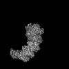
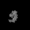
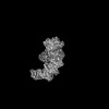
| Entry | Database: PDB / ID: 7xf1 | ||||||
|---|---|---|---|---|---|---|---|
| Title | Crystal strucutre of apoCasDinG in complex with ssDNA | ||||||
 Components Components |
| ||||||
 Keywords Keywords | STRUCTURAL PROTEIN/DNA / Nuclease / STRUCTURAL PROTEIN / STRUCTURAL PROTEIN-DNA complex | ||||||
| Function / homology | P-loop containing nucleotide triphosphate hydrolases / Rossmann fold / 3-Layer(aba) Sandwich / Alpha Beta / DNA / DNA (> 10) Function and homology information Function and homology information | ||||||
| Biological species |   Pseudomonas aeruginosa (bacteria) Pseudomonas aeruginosa (bacteria)synthetic construct (others) | ||||||
| Method |  X-RAY DIFFRACTION / SYNCHROTRON / MOLECULAR REPLACEMENT / Resolution: 3.2 Å X-RAY DIFFRACTION / SYNCHROTRON / MOLECULAR REPLACEMENT / Resolution: 3.2 Å | ||||||
 Authors Authors | Zhang, J.T. / Cui, N. / Liu, Y.R. / Huang, H.D. / Jia, N. | ||||||
| Funding support | 1items
| ||||||
 Citation Citation | Journal: Mol Cell / Year: 2023 Title: Type IV-A CRISPR-Csf complex: Assembly, dsDNA targeting, and CasDinG recruitment. Authors: Ning Cui / Jun-Tao Zhang / Yongrui Liu / Yanhong Liu / Xiao-Yu Liu / Chongyuan Wang / Hongda Huang / Ning Jia /  Abstract: Type IV CRISPR-Cas systems, which are primarily found on plasmids and exhibit a strong plasmid-targeting preference, are the only one of the six known CRISPR-Cas types for which the mechanistic ...Type IV CRISPR-Cas systems, which are primarily found on plasmids and exhibit a strong plasmid-targeting preference, are the only one of the six known CRISPR-Cas types for which the mechanistic details of their function remain unknown. Here, we provide high-resolution functional snapshots of type IV-A Csf complexes before and after target dsDNA binding, either in the absence or presence of CasDinG, revealing the mechanisms underlying Csf complex assembly, "DWN" PAM-dependent dsDNA targeting, R-loop formation, and CasDinG recruitment. Furthermore, we establish that CasDinG, a signature DinG family helicase, harbors ssDNA-stimulated ATPase activity and ATP-dependent 5'-3' DNA helicase activity. In addition, we show that CasDinG unwinds the non-target strand (NTS) and target strand (TS) of target dsDNA from the Csf complex. These molecular details advance our mechanistic understanding of type IV-A CRISPR-Csf function and should enable Csf complexes to be harnessed as genome-engineering tools for biotechnological applications. | ||||||
| History |
|
- Structure visualization
| Structure viewer | Molecule: MolmilJmol/JSmol |
|---|
- Downloads & links
-Download
| PDBx/mmCIF format | 7xf1.cif.gz | 248.5 KB | Display | PDBx/mmCIF format |
|---|---|---|---|---|
| PDB format | pdb7xf1.ent.gz | 200.3 KB | Display | PDB format |
| PDBx/mmJSON format | 7xf1.json.gz | Tree view | PDBx/mmJSON format | |
| Others |  Other downloads Other downloads |
-Validation report
| Arichive directory | https://data.pdbj.org/pub/pdb/validation_reports/xf/7xf1ftp://data.pdbj.org/pub/pdb/validation_reports/xf/7xf1 | HTTPS FTP |
|---|
-Related structure data
| Related structure data | 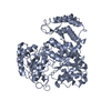 7xexC  7xf0C  7xfzC  7xg0C  7xg2C  7xg3C  7xg4C 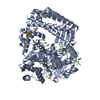 6fwrS S: Starting model for refinement C: citing same article ( |
|---|---|
| Similar structure data |
-Links
 PDBj
PDBj


- Assembly
| Deposited unit | 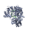
| ||||||||
|---|---|---|---|---|---|---|---|---|---|
| 1 |
| ||||||||
| Unit cell |
|
-Components
| #1: Protein | Mass: 67939.883 Da / Num. of mol.: 1 Source method: isolated from a genetically manipulated source Details: Sequence based on database WP_088922490.1 / Source: (gene. exp.) Pseudomonas aeruginosa (bacteria) / Production host: |
|---|---|
| #2: DNA chain | Mass: 3301.163 Da / Num. of mol.: 1 / Source method: obtained synthetically / Details: ssDNA12 / Source: (synth.) synthetic construct (others) |
| Has protein modification | N |
-Experimental details
-Experiment
| Experiment | Method: X-RAY DIFFRACTION / Number of used crystals: 1 |
|---|
- Sample preparation
| Crystal | Density Matthews: 3.83 Å3/Da / Density % sol: 67.84 % |
|---|---|
| Crystal grow | Temperature: 289.15 K / Method: vapor diffusion, hanging drop / Details: 0.1M MES pH 6.0, 1.6M Ammonium sulfate |
-Data collection
| Diffraction | Mean temperature: 100 K / Serial crystal experiment: N |
|---|---|
| Diffraction source | Source: SYNCHROTRON / Site: SSRF / Beamline: BL17B1 / Wavelength: 0.9792 Å |
| Detector | Type: DECTRIS EIGER X 16M / Detector: PIXEL / Date: Nov 7, 2021 |
| Radiation | Protocol: SINGLE WAVELENGTH / Monochromatic (M) / Laue (L): M / Scattering type: x-ray |
| Radiation wavelength | Wavelength: 0.9792 Å / Relative weight: 1 |
| Reflection | Resolution: 3.2→40 Å / Num. obs: 18489 / % possible obs: 99.8 % / Redundancy: 18 % / Biso Wilson estimate: 67.91 Å2 / CC1/2: 0.984 / Net I/σ(I): 7 |
| Reflection shell | Resolution: 3.2→3.31 Å / Num. unique obs: 1797 / CC1/2: 0.575 |
- Processing
| Software |
| |||||||||||||||||||||||||||||||||||||||||||||||||||||||||||||||||||||||||||
|---|---|---|---|---|---|---|---|---|---|---|---|---|---|---|---|---|---|---|---|---|---|---|---|---|---|---|---|---|---|---|---|---|---|---|---|---|---|---|---|---|---|---|---|---|---|---|---|---|---|---|---|---|---|---|---|---|---|---|---|---|---|---|---|---|---|---|---|---|---|---|---|---|---|---|---|---|
| Refinement | Method to determine structure: MOLECULAR REPLACEMENT Starting model: 6FWR Resolution: 3.2→25.46 Å / SU ML: 0.34 / Cross valid method: THROUGHOUT / σ(F): 1.35 / Phase error: 20.63 / Stereochemistry target values: ML
| |||||||||||||||||||||||||||||||||||||||||||||||||||||||||||||||||||||||||||
| Solvent computation | Shrinkage radii: 0.9 Å / VDW probe radii: 1.11 Å / Solvent model: FLAT BULK SOLVENT MODEL | |||||||||||||||||||||||||||||||||||||||||||||||||||||||||||||||||||||||||||
| Displacement parameters | Biso max: 152.81 Å2 / Biso mean: 66.265 Å2 / Biso min: 29.25 Å2 | |||||||||||||||||||||||||||||||||||||||||||||||||||||||||||||||||||||||||||
| Refinement step | Cycle: final / Resolution: 3.2→25.46 Å
| |||||||||||||||||||||||||||||||||||||||||||||||||||||||||||||||||||||||||||
| LS refinement shell | Refine-ID: X-RAY DIFFRACTION / Rfactor Rfree error: 0 / Total num. of bins used: 6
| |||||||||||||||||||||||||||||||||||||||||||||||||||||||||||||||||||||||||||
| Refinement TLS params. | Method: refined / Refine-ID: X-RAY DIFFRACTION
| |||||||||||||||||||||||||||||||||||||||||||||||||||||||||||||||||||||||||||
| Refinement TLS group |
|
