 Movie
Movie Controller

[English] 日本語
 Yorodumi
- PDB-7xfh: Structure of nucleosome-AAG complex (A-30I, post-catalytic state) -
+ Open data

- Basic information
| Entry | Database: PDB / ID: 7xfh | ||||||
|---|---|---|---|---|---|---|---|
| Title | Structure of nucleosome-AAG complex (A-30I, post-catalytic state) | ||||||
 Components Components |
| ||||||
 Keywords Keywords | DNA BINDING PROTEIN/DNA / Nucleosome / AAG / DNA repair / Base excision repair / DNA BINDING PROTEIN / DNA BINDING PROTEIN-DNA complex | ||||||
| Function / homology |  Function and homology information Function and homology informationalkylbase DNA N-glycosylase activity / DNA-7-methyladenine glycosylase activity / DNA-3-methylguanine glycosylase activity / DNA-3-methyladenine glycosylase II / DNA-7-methylguanine glycosylase activity / DNA-3-methyladenine glycosylase activity / depurination / DNA N-glycosylase activity / Displacement of DNA glycosylase by APEX1 / DNA alkylation repair ...alkylbase DNA N-glycosylase activity / DNA-7-methyladenine glycosylase activity / DNA-3-methylguanine glycosylase activity / DNA-3-methyladenine glycosylase II / DNA-7-methylguanine glycosylase activity / DNA-3-methyladenine glycosylase activity / depurination / DNA N-glycosylase activity / Displacement of DNA glycosylase by APEX1 / DNA alkylation repair / mitochondrial nucleoid / Recognition and association of DNA glycosylase with site containing an affected purine / Cleavage of the damaged purine / base-excision repair / structural constituent of chromatin / nucleosome / nucleosome assembly / damaged DNA binding / protein heterodimerization activity / DNA binding / nucleoplasm / nucleus / cytosol Similarity search - Function | ||||||
| Biological species |  Homo sapiens (human) Homo sapiens (human) | ||||||
| Method | ELECTRON MICROSCOPY / single particle reconstruction / negative staining / cryo EM / Resolution: 2.9 Å | ||||||
 Authors Authors | Zheng, L. / Tsai, B. / Gao, N. | ||||||
| Funding support |  China, 1items China, 1items
| ||||||
 Citation Citation | Journal: Cell Discov / Year: 2023 Title: Structural and mechanistic insights into the DNA glycosylase AAG-mediated base excision in nucleosome. Authors: Lvqin Zheng / Bin Tsai / Ning Gao / Abstract: The engagement of a DNA glycosylase with a damaged DNA base marks the initiation of base excision repair. Nucleosome-based packaging of eukaryotic genome obstructs DNA accessibility, and how DNA ...The engagement of a DNA glycosylase with a damaged DNA base marks the initiation of base excision repair. Nucleosome-based packaging of eukaryotic genome obstructs DNA accessibility, and how DNA glycosylases locate the substrate site on nucleosomes is currently unclear. Here, we report cryo-electron microscopy structures of nucleosomes bearing a deoxyinosine (DI) in various geometric positions and structures of them in complex with the DNA glycosylase AAG. The apo nucleosome structures show that the presence of a DI alone perturbs nucleosomal DNA globally, leading to a general weakening of the interface between DNA and the histone core and greater flexibility for the exit/entry of the nucleosomal DNA. AAG makes use of this nucleosomal plasticity and imposes further local deformation of the DNA through formation of the stable enzyme-substrate complex. Mechanistically, local distortion augmentation, translation/rotational register shift and partial opening of the nucleosome are employed by AAG to cope with substrate sites in fully exposed, occluded and completely buried positions, respectively. Our findings reveal the molecular basis for the DI-induced modification on the structural dynamics of the nucleosome and elucidate how the DNA glycosylase AAG accesses damaged sites on the nucleosome with different solution accessibility. | ||||||
| History |
|
- Structure visualization
| Structure viewer | Molecule: MolmilJmol/JSmol |
|---|
- Downloads & links
-Download
| PDBx/mmCIF format | 7xfh.cif.gz | 309.6 KB | Display | PDBx/mmCIF format |
|---|---|---|---|---|
| PDB format | pdb7xfh.ent.gz | 231.4 KB | Display | PDB format |
| PDBx/mmJSON format | 7xfh.json.gz | Tree view | PDBx/mmJSON format | |
| Others |  Other downloads Other downloads |
-Validation report
| Summary document | 7xfh_validation.pdf.gz | 1.1 MB | Display | wwPDB validaton report |
|---|---|---|---|---|
| Full document | 7xfh_full_validation.pdf.gz | 1.1 MB | Display | |
| Data in XML | 7xfh_validation.xml.gz | 37.1 KB | Display | |
| Data in CIF | 7xfh_validation.cif.gz | 57.6 KB | Display | |
| Arichive directory | https://data.pdbj.org/pub/pdb/validation_reports/xf/7xfhftp://data.pdbj.org/pub/pdb/validation_reports/xf/7xfh | HTTPS FTP |
-Related structure data
| Related structure data |  33172MC 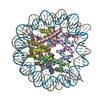 7xfcC 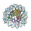 7xfiC  7xfjC 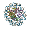 7xflC  7xfmC 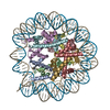 7xfnC 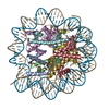 7xnpC M: map data used to model this data C: citing same article ( |
|---|---|
| Similar structure data |

-Links
 PDBj
PDBj


- Assembly
| Deposited unit | 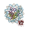
|
|---|---|
| 1 |
|
-Components
-Protein , 5 types, 9 molecules AEBFCGDHK
| #1: Protein | Mass: 15421.101 Da / Num. of mol.: 2 Source method: isolated from a genetically manipulated source Source: (gene. exp.)  #2: Protein | Mass: 11394.426 Da / Num. of mol.: 2 Source method: isolated from a genetically manipulated source Source: (gene. exp.) #3: Protein | Mass: 14093.436 Da / Num. of mol.: 2 Source method: isolated from a genetically manipulated source Source: (gene. exp.) #4: Protein | Mass: 13965.265 Da / Num. of mol.: 2 Source method: isolated from a genetically manipulated source Source: (gene. exp.) #7: Protein | | Mass: 32915.699 Da / Num. of mol.: 1 Source method: isolated from a genetically manipulated source Source: (gene. exp.) Homo sapiens (human) / Gene: MPG, AAG, ANPG, MID1 / Production host: References: UniProt: P29372, DNA-3-methyladenine glycosylase II |
|---|
-DNA chain , 2 types, 2 molecules IJ
| #5: DNA chain | Mass: 47051.930 Da / Num. of mol.: 1 Source method: isolated from a genetically manipulated source Source: (gene. exp.) |
|---|---|
| #6: DNA chain | Mass: 46655.711 Da / Num. of mol.: 1 Source method: isolated from a genetically manipulated source Source: (gene. exp.) |
-Details
| Has ligand of interest | Y |
|---|
-Experimental details
-Experiment
| Experiment | Method: ELECTRON MICROSCOPY |
|---|---|
| EM experiment | Aggregation state: CELL / 3D reconstruction method: single particle reconstruction |
- Sample preparation
| Component |
| ||||||||||||||||||||||||
|---|---|---|---|---|---|---|---|---|---|---|---|---|---|---|---|---|---|---|---|---|---|---|---|---|---|
| Source (natural) |
| ||||||||||||||||||||||||
| Source (recombinant) |
| ||||||||||||||||||||||||
| Buffer solution | pH: 7.5 | ||||||||||||||||||||||||
| Specimen | Embedding applied: NO / Shadowing applied: NO / Staining applied: YES / Vitrification applied: YES | ||||||||||||||||||||||||
| EM staining | Type: NEGATIVE / Material: Uranyl Acetate | ||||||||||||||||||||||||
| Vitrification | Cryogen name: NITROGEN |
- Electron microscopy imaging
| Experimental equipment |  Model: Titan Krios / Image courtesy: FEI Company |
|---|---|
| Microscopy | Model: FEI TITAN KRIOS |
| Electron gun | Electron source:  FIELD EMISSION GUN / Accelerating voltage: 300 kV / Illumination mode: SPOT SCAN FIELD EMISSION GUN / Accelerating voltage: 300 kV / Illumination mode: SPOT SCAN |
| Electron lens | Mode: DARK FIELD / Nominal defocus max: 1700 nm / Nominal defocus min: 1200 nm |
| Image recording | Electron dose: 64 e/Å2 / Film or detector model: GATAN K2 SUMMIT (4k x 4k) |
- Processing
| Software | Name: PHENIX / Version: 1.19.2_4158: / Classification: refinement | ||||||||||||||||||||||||
|---|---|---|---|---|---|---|---|---|---|---|---|---|---|---|---|---|---|---|---|---|---|---|---|---|---|
| CTF correction | Type: PHASE FLIPPING AND AMPLITUDE CORRECTION | ||||||||||||||||||||||||
| 3D reconstruction | Resolution: 2.9 Å / Resolution method: FSC 0.143 CUT-OFF / Num. of particles: 126881 / Symmetry type: POINT | ||||||||||||||||||||||||
| Refine LS restraints |
|
