 Movie
Movie Controller


+ Open data

- Basic information
| Entry | Database: PDB / ID: 7xdi | ||||||||||||||||||
|---|---|---|---|---|---|---|---|---|---|---|---|---|---|---|---|---|---|---|---|
| Title | Tail structure of bacteriophage SSV19 | ||||||||||||||||||
 Components Components |
| ||||||||||||||||||
 Keywords Keywords | VIRAL PROTEIN / Complex | ||||||||||||||||||
| Function / homology |  Function and homology information Function and homology informationcarbohydrate metabolic process / structural molecule activity / membrane Similarity search - Function | ||||||||||||||||||
| Biological species |   Sulfolobus spindle-shaped virus Sulfolobus spindle-shaped virus | ||||||||||||||||||
| Method | ELECTRON MICROSCOPY / single particle reconstruction / cryo EM / Resolution: 3.8 Å | ||||||||||||||||||
 Authors Authors | Liu, H.R. / Chen, W.Y. | ||||||||||||||||||
| Funding support |  China, 5items China, 5items
| ||||||||||||||||||
 Citation Citation | Journal: Proc Natl Acad Sci U S A / Year: 2022 Title: Structural insights into a spindle-shaped archaeal virus with a sevenfold symmetrical tail. Authors: Zhen Han / Wanjuan Yuan / Hao Xiao / Li Wang / Junxia Zhang / Yuning Peng / Lingpeng Cheng / Hongrong Liu / Li Huang / Abstract: Archaeal viruses with a spindle-shaped virion are abundant and widespread in extremely diverse environments. However, efforts to obtain the high-resolution structure of a spindle-shaped virus have ...Archaeal viruses with a spindle-shaped virion are abundant and widespread in extremely diverse environments. However, efforts to obtain the high-resolution structure of a spindle-shaped virus have been unsuccessful. Here, we present the structure of SSV19, a spindle-shaped virus infecting the hyperthermophilic archaeon sp. E11-6. Our near-atomic structure reveals an unusual sevenfold symmetrical virus tail consisting of the tailspike, nozzle, and adaptor proteins. The spindle-shaped capsid shell is formed by seven left-handed helical strands, constructed of the hydrophobic major capsid protein, emanating from the highly glycosylated tail assembly. Sliding between adjacent strands is responsible for the variation of a virion in size. Ultrathin sections of the SSV19-infected cells show that SSV19 virions adsorb to the host cell membrane through the tail after penetrating the S-layer. The tailspike harbors a putative endo-mannanase domain, which shares structural similarity to a endo-mannanase. Molecules of glycerol dibiphytanyl glycerol tetraether lipid were observed in hydrophobic clefts between the tail and the capsid shell. The nozzle protein resembles the stem and clip domains of the portals of herpesviruses and bacteriophages, implying an evolutionary relationship among the archaeal, bacterial, and eukaryotic viruses. | ||||||||||||||||||
| History |
|
- Structure visualization
| Structure viewer | Molecule: MolmilJmol/JSmol |
|---|
- Downloads & links
-Download
| PDBx/mmCIF format | 7xdi.cif.gz | 194.4 KB | Display | PDBx/mmCIF format |
|---|---|---|---|---|
| PDB format | pdb7xdi.ent.gz | 144.3 KB | Display | PDB format |
| PDBx/mmJSON format | 7xdi.json.gz | Tree view | PDBx/mmJSON format | |
| Others |  Other downloads Other downloads |
-Validation report
| Summary document | 7xdi_validation.pdf.gz | 1.2 MB | Display | wwPDB validaton report |
|---|---|---|---|---|
| Full document | 7xdi_full_validation.pdf.gz | 1.2 MB | Display | |
| Data in XML | 7xdi_validation.xml.gz | 41 KB | Display | |
| Data in CIF | 7xdi_validation.cif.gz | 58.4 KB | Display | |
| Arichive directory | https://data.pdbj.org/pub/pdb/validation_reports/xd/7xdiftp://data.pdbj.org/pub/pdb/validation_reports/xd/7xdi | HTTPS FTP |
-Related structure data
| Related structure data | 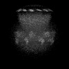 33148MC M: map data used to model this data C: citing same article ( |
|---|---|
| Similar structure data |

-Links
 PDBj
PDBj
- Assembly
| Deposited unit | 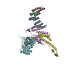
|
|---|---|
| 1 | x 7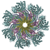
|
| 2 |
|
| 3 | 
|
| Symmetry | Point symmetry: (Schoenflies symbol: C7 (7 fold cyclic)) |
-Components
| #1: Protein | Mass: 9282.206 Da / Num. of mol.: 3 / Source method: isolated from a natural source / Source: (natural) Sulfolobus spindle-shaped virus / References: UniProt: A0A5Q0V137#2: Protein | | Mass: 14753.507 Da / Num. of mol.: 1 / Source method: isolated from a natural source / Source: (natural) Sulfolobus spindle-shaped virus / References: UniProt: A0A5Q0V0G9#3: Protein | | Mass: 23195.996 Da / Num. of mol.: 1 / Source method: isolated from a natural source / Source: (natural) Sulfolobus spindle-shaped virus / References: UniProt: A0A5Q0V0F6#4: Protein | | Mass: 137071.953 Da / Num. of mol.: 1 / Source method: isolated from a natural source / Source: (natural) Sulfolobus spindle-shaped virus / References: UniProt: A0A5Q0V0A2 |
|---|
-Experimental details
-Experiment
| Experiment | Method: ELECTRON MICROSCOPY |
|---|---|
| EM experiment | Aggregation state: PARTICLE / 3D reconstruction method: single particle reconstruction |
- Sample preparation
| Component | Name: Sulfolobus spindle-shaped virus / Type: VIRUS / Entity ID: all / Source: NATURAL |
|---|---|
| Source (natural) | Organism: Sulfolobus spindle-shaped virus |
| Details of virus | Empty: NO / Enveloped: NO / Isolate: OTHER / Type: VIRION |
| Buffer solution | pH: 7 |
| Specimen | Embedding applied: NO / Shadowing applied: NO / Staining applied: NO / Vitrification applied: YES |
| Vitrification | Cryogen name: ETHANE |
- Electron microscopy imaging
| Experimental equipment |  Model: Talos Arctica / Image courtesy: FEI Company |
|---|---|
| Microscopy | Model: FEI TECNAI ARCTICA |
| Electron gun | Electron source:  FIELD EMISSION GUN / Accelerating voltage: 200 kV / Illumination mode: SPOT SCAN FIELD EMISSION GUN / Accelerating voltage: 200 kV / Illumination mode: SPOT SCAN |
| Electron lens | Mode: BRIGHT FIELD / Nominal defocus max: 4000 nm / Nominal defocus min: 500 nm |
| Image recording | Electron dose: 35 e/Å2 / Film or detector model: FEI FALCON II (4k x 4k) |
- Processing
| Software | Name: PHENIX / Version: 1.20.1_4487: / Classification: refinement | ||||||||||||||||||||||||
|---|---|---|---|---|---|---|---|---|---|---|---|---|---|---|---|---|---|---|---|---|---|---|---|---|---|
| CTF correction | Type: NONE | ||||||||||||||||||||||||
| 3D reconstruction | Resolution: 3.8 Å / Resolution method: FSC 0.143 CUT-OFF / Num. of particles: 49361 / Symmetry type: POINT | ||||||||||||||||||||||||
| Refine LS restraints |
|
