 Movie
Movie Controller

[English] 日本語
 Yorodumi
- PDB-7wlz: Cryo-EM structure of the Omicron S in complex with 35B5 Fab(1 dow... -
+ Open data

- Basic information
| Entry | Database: PDB / ID: 7wlz | |||||||||
|---|---|---|---|---|---|---|---|---|---|---|
| Title | Cryo-EM structure of the Omicron S in complex with 35B5 Fab(1 down-, 1 up- and 1 invisible RBDs) | |||||||||
 Components Components |
| |||||||||
 Keywords Keywords | VIRAL PROTEIN / IMMUNE SYSTEM | |||||||||
| Function / homology |  Function and homology information Function and homology informationMaturation of spike protein / viral translation / Translation of Structural Proteins / Virion Assembly and Release / host cell surface / host extracellular space / suppression by virus of host tetherin activity / Induction of Cell-Cell Fusion / structural constituent of virion / entry receptor-mediated virion attachment to host cell ...Maturation of spike protein / viral translation / Translation of Structural Proteins / Virion Assembly and Release / host cell surface / host extracellular space / suppression by virus of host tetherin activity / Induction of Cell-Cell Fusion / structural constituent of virion / entry receptor-mediated virion attachment to host cell / host cell endoplasmic reticulum-Golgi intermediate compartment membrane / membrane fusion / receptor-mediated endocytosis of virus by host cell / Attachment and Entry / positive regulation of viral entry into host cell / receptor-mediated virion attachment to host cell / receptor ligand activity / symbiont-mediated suppression of host innate immune response / host cell surface receptor binding / fusion of virus membrane with host plasma membrane / fusion of virus membrane with host endosome membrane / viral envelope / virion attachment to host cell / SARS-CoV-2 activates/modulates innate and adaptive immune responses / host cell plasma membrane / virion membrane / identical protein binding / membrane / plasma membrane Similarity search - Function | |||||||||
| Biological species |   Severe acute respiratory syndrome coronavirus 2 Severe acute respiratory syndrome coronavirus 2 Homo sapiens (human) Homo sapiens (human) | |||||||||
| Method | ELECTRON MICROSCOPY / single particle reconstruction / cryo EM / Resolution: 2.98 Å | |||||||||
 Authors Authors | Wang, X. / Zhu, Y. | |||||||||
| Funding support |  China, 2items China, 2items
| |||||||||
 Citation Citation | Journal: Cell Host Microbe / Year: 2022 Title: 35B5 antibody potently neutralizes SARS-CoV-2 Omicron by disrupting the N-glycan switch via a conserved spike epitope. Authors: Xiaofei Wang / Xiangyu Chen / Jiaxing Tan / Shuai Yue / Runhong Zhou / Yan Xu / Yao Lin / Yang Yang / Yan Zhou / Kai Deng / Zhiwei Chen / Lilin Ye / Yongqun Zhu / Abstract: The SARS-CoV-2 Omicron variant harbors more than 30 mutations in the spike protein, leading to immune evasion from many therapeutic neutralizing antibodies. We reveal that a receptor-binding domain ...The SARS-CoV-2 Omicron variant harbors more than 30 mutations in the spike protein, leading to immune evasion from many therapeutic neutralizing antibodies. We reveal that a receptor-binding domain (RBD)-targeting monoclonal antibody, 35B5, exhibits potent neutralizing efficacy to Omicron. Cryo-electron microscopy structures of the extracellular domain trimer of Omicron spike with 35B5 Fab reveal that Omicron spike exhibits tight trimeric packing and high thermostability, as well as significant antigenic shifts and structural changes, within the RBD, N-terminal domain (NTD), and subdomains 1 and 2. However, these changes do not affect targeting of the invariant 35B5 epitope. 35B5 potently neutralizes SARS-CoV-2 Omicron and other variants by causing significant conformational changes within a conserved N-glycan switch that controls the transition of RBD from the "down" state to the "up" state, which allows recognition of the host entry receptor ACE2. This mode of action and potent neutralizing capacity of 35B5 indicate its potential therapeutic application for SARS-CoV-2. | |||||||||
| History |
|
- Structure visualization
| Structure viewer | Molecule: MolmilJmol/JSmol |
|---|
- Downloads & links
-Download
| PDBx/mmCIF format | 7wlz.cif.gz | 575.5 KB | Display | PDBx/mmCIF format |
|---|---|---|---|---|
| PDB format | pdb7wlz.ent.gz | 461.4 KB | Display | PDB format |
| PDBx/mmJSON format | 7wlz.json.gz | Tree view | PDBx/mmJSON format | |
| Others |  Other downloads Other downloads |
-Validation report
| Summary document | 7wlz_validation.pdf.gz | 899.9 KB | Display | wwPDB validaton report |
|---|---|---|---|---|
| Full document | 7wlz_full_validation.pdf.gz | 945.5 KB | Display | |
| Data in XML | 7wlz_validation.xml.gz | 86.1 KB | Display | |
| Data in CIF | 7wlz_validation.cif.gz | 131.7 KB | Display | |
| Arichive directory | https://data.pdbj.org/pub/pdb/validation_reports/wl/7wlzftp://data.pdbj.org/pub/pdb/validation_reports/wl/7wlz | HTTPS FTP |
-Related structure data
| Related structure data |  32595MC 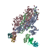 7wlyC M: map data used to model this data C: citing same article ( |
|---|---|
| Similar structure data |

-Links
 PDBj
PDBj


- Assembly
| Deposited unit | 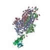
|
|---|---|
| 1 |
|
-Components
| #1: Protein | Mass: 142586.156 Da / Num. of mol.: 3 Source method: isolated from a genetically manipulated source Source: (gene. exp.) Severe acute respiratory syndrome coronavirus 2Gene: S, 2 / Variant: Omicron / Cell line (production host): HEK293 / Production host: Homo sapiens (human) / References: UniProt: P0DTC2#2: Antibody | | Mass: 25139.314 Da / Num. of mol.: 1 Source method: isolated from a genetically manipulated source Source: (gene. exp.) Homo sapiens (human) / Cell line (production host): HEK293 / Production host: Homo sapiens (human)#3: Antibody | | Mass: 23905.633 Da / Num. of mol.: 1 Source method: isolated from a genetically manipulated source Source: (gene. exp.) Homo sapiens (human) / Cell line (production host): HEK293 / Production host: Homo sapiens (human)#4: Sugar | ChemComp-NAG /   Type: D-saccharide, beta linking / Mass: 221.208 Da / Num. of mol.: 27 / Source method: obtained synthetically / Formula: C8H15NO6 Type: D-saccharide, beta linking / Mass: 221.208 Da / Num. of mol.: 27 / Source method: obtained synthetically / Formula: C8H15NO6Has ligand of interest | N | |
|---|
-Experimental details
-Experiment
| Experiment | Method: ELECTRON MICROSCOPY |
|---|---|
| EM experiment | Aggregation state: PARTICLE / 3D reconstruction method: single particle reconstruction |
- Sample preparation
| Component |
| ||||||||||||||||||||||||
|---|---|---|---|---|---|---|---|---|---|---|---|---|---|---|---|---|---|---|---|---|---|---|---|---|---|
| Molecular weight | Experimental value: NO | ||||||||||||||||||||||||
| Source (natural) |
| ||||||||||||||||||||||||
| Source (recombinant) |
| ||||||||||||||||||||||||
| Buffer solution | pH: 7.2 | ||||||||||||||||||||||||
| Specimen | Embedding applied: NO / Shadowing applied: NO / Staining applied: NO / Vitrification applied: YES | ||||||||||||||||||||||||
| Specimen support | Grid material: GOLD / Grid mesh size: 300 divisions/in. / Grid type: Quantifoil R1.2/1.3 | ||||||||||||||||||||||||
| Vitrification | Cryogen name: ETHANE |
- Electron microscopy imaging
| Experimental equipment |  Model: Titan Krios / Image courtesy: FEI Company |
|---|---|
| Microscopy | Model: FEI TITAN KRIOS |
| Electron gun | Electron source:  FIELD EMISSION GUN / Accelerating voltage: 300 kV / Illumination mode: SPOT SCAN FIELD EMISSION GUN / Accelerating voltage: 300 kV / Illumination mode: SPOT SCAN |
| Electron lens | Mode: BRIGHT FIELD / Nominal defocus max: 1300 nm / Nominal defocus min: 1000 nm |
| Image recording | Electron dose: 64 e/Å2 / Film or detector model: GATAN K2 SUMMIT (4k x 4k) |
- Processing
| Software | Name: PHENIX / Version: 1.18.2_3874: / Classification: refinement | ||||||||||||||||||||||||
|---|---|---|---|---|---|---|---|---|---|---|---|---|---|---|---|---|---|---|---|---|---|---|---|---|---|
| EM software |
| ||||||||||||||||||||||||
| CTF correction | Type: PHASE FLIPPING AND AMPLITUDE CORRECTION | ||||||||||||||||||||||||
| Symmetry | Point symmetry: C1 (asymmetric) | ||||||||||||||||||||||||
| 3D reconstruction | Resolution: 2.98 Å / Resolution method: FSC 0.143 CUT-OFF / Num. of particles: 243427 / Symmetry type: POINT | ||||||||||||||||||||||||
| Refine LS restraints |
|
