[English] 日本語
 Yorodumi
- PDB-7wlj: CryoEM structure of human low-voltage activated T-type calcium ch... -
+ Open data
- Basic information
| Entry | Database: PDB / ID: 7wlj | ||||||
|---|---|---|---|---|---|---|---|
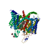
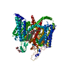
| Title | CryoEM structure of human low-voltage activated T-type calcium channel Cav3.3 in complex with mibefradil (MIB) | ||||||
 Components Components | Voltage-dependent T-type calcium channel subunit alpha-1I | ||||||
 Keywords Keywords | MEMBRANE PROTEIN / MIB | ||||||
| Function / homology |  Function and homology information Function and homology informationhigh voltage-gated calcium channel activity / sleep / NCAM1 interactions / voltage-gated calcium channel complex / calcium ion import across plasma membrane / neuronal action potential / Smooth Muscle Contraction / voltage-gated calcium channel activity / signal transduction / plasma membrane Similarity search - Function | ||||||
| Biological species |  Homo sapiens (human) Homo sapiens (human) | ||||||
| Method | ELECTRON MICROSCOPY / single particle reconstruction / cryo EM / Resolution: 3.9 Å | ||||||
 Authors Authors | He, L. / Yu, Z. / Dong, Y. / Chen, Q. / Zhao, Y. | ||||||
| Funding support |  China, 1items China, 1items
| ||||||
 Citation Citation |  Journal: Nat Commun / Year: 2022 Journal: Nat Commun / Year: 2022Title: Structure, gating, and pharmacology of human Ca3.3 channel. Authors: Lingli He / Zhuoya Yu / Ze Geng / Zhuo Huang / Changjiang Zhang / Yanli Dong / Yiwei Gao / Yuhang Wang / Qihao Chen / Le Sun / Xinyue Ma / Bo Huang / Xiaoqun Wang / Yan Zhao /  Abstract: The low-voltage activated T-type calcium channels regulate cellular excitability and oscillatory behavior of resting membrane potential which trigger many physiological events and have been ...The low-voltage activated T-type calcium channels regulate cellular excitability and oscillatory behavior of resting membrane potential which trigger many physiological events and have been implicated with many diseases. Here, we determine structures of the human T-type Ca3.3 channel, in the absence and presence of antihypertensive drug mibefradil, antispasmodic drug otilonium bromide and antipsychotic drug pimozide. Ca3.3 contains a long bended S6 helix from domain III, with a positive charged region protruding into the cytosol, which is critical for T-type Ca channel activation at low voltage. The drug-bound structures clearly illustrate how these structurally different compounds bind to the same central cavity inside the Ca3.3 channel, but are mediated by significantly distinct interactions between drugs and their surrounding residues. Phospholipid molecules penetrate into the central cavity in various extent to shape the binding pocket and play important roles in stabilizing the inhibitor. These structures elucidate mechanisms of channel gating, drug recognition, and actions, thus pointing the way to developing potent and subtype-specific drug for therapeutic treatments of related disorders. | ||||||
| History |
|
- Structure visualization
| Structure viewer | Molecule:  Molmil Molmil Jmol/JSmol Jmol/JSmol |
|---|
- Downloads & links
- Download
| PDBx/mmCIF format |  7wlj.cif.gz 7wlj.cif.gz | 238.8 KB | Display |  PDBx/mmCIF format PDBx/mmCIF format |
|---|---|---|---|---|
| PDB format |  pdb7wlj.ent.gz pdb7wlj.ent.gz | 183.7 KB | Display |  PDB format PDB format |
| PDBx/mmJSON format |  7wlj.json.gz 7wlj.json.gz | Tree view |  PDBx/mmJSON format PDBx/mmJSON format | |
| Others |  Other downloads Other downloads |
-Validation report
| Summary document |  7wlj_validation.pdf.gz 7wlj_validation.pdf.gz | 1.3 MB | Display |  wwPDB validaton report wwPDB validaton report |
|---|---|---|---|---|
| Full document |  7wlj_full_validation.pdf.gz 7wlj_full_validation.pdf.gz | 1.3 MB | Display | |
| Data in XML |  7wlj_validation.xml.gz 7wlj_validation.xml.gz | 43 KB | Display | |
| Data in CIF |  7wlj_validation.cif.gz 7wlj_validation.cif.gz | 62.5 KB | Display | |
| Arichive directory |  https://data.pdbj.org/pub/pdb/validation_reports/wl/7wlj https://data.pdbj.org/pub/pdb/validation_reports/wl/7wlj ftp://data.pdbj.org/pub/pdb/validation_reports/wl/7wlj ftp://data.pdbj.org/pub/pdb/validation_reports/wl/7wlj | HTTPS FTP |
-Related structure data
| Related structure data |  32585MC  7wliC  7wlkC  7wllC M: map data used to model this data C: citing same article ( |
|---|---|
| Similar structure data | Similarity search - Function & homology  F&H Search F&H Search |
- Links
Links
- Assembly
| Deposited unit | 
|
|---|---|
| 1 |
|
- Components
-Protein / Sugars , 2 types, 3 molecules A

| #1: Protein | Mass: 245373.031 Da / Num. of mol.: 1 Source method: isolated from a genetically manipulated source Source: (gene. exp.)  Homo sapiens (human) / Gene: CACNA1I, KIAA1120 / Production host: Homo sapiens (human) / Gene: CACNA1I, KIAA1120 / Production host:  Homo sapiens (human) / References: UniProt: Q9P0X4 Homo sapiens (human) / References: UniProt: Q9P0X4 |
|---|---|
| #6: Sugar |
-Non-polymers , 4 types, 13 molecules 


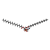
| #2: Chemical | ChemComp-MWV / ( | ||||
|---|---|---|---|---|---|
| #3: Chemical | ChemComp-3PE / #4: Chemical | ChemComp-CA / | #5: Chemical | ChemComp-Y01 / |
-Details
| Has ligand of interest | Y |
|---|
-Experimental details
-Experiment
| Experiment | Method: ELECTRON MICROSCOPY |
|---|---|
| EM experiment | Aggregation state: PARTICLE / 3D reconstruction method: single particle reconstruction |
- Sample preparation
| Component | Name: CaV3.3 / Type: COMPLEX / Entity ID: #1 / Source: RECOMBINANT |
|---|---|
| Molecular weight | Experimental value: NO |
| Source (natural) | Organism:  Homo sapiens (human) Homo sapiens (human) |
| Source (recombinant) | Organism:  Homo sapiens (human) Homo sapiens (human) |
| Buffer solution | pH: 7.5 |
| Specimen | Embedding applied: NO / Shadowing applied: NO / Staining applied: NO / Vitrification applied: YES |
| Vitrification | Cryogen name: ETHANE |
- Electron microscopy imaging
| Experimental equipment |  Model: Titan Krios / Image courtesy: FEI Company |
|---|---|
| Microscopy | Model: FEI TITAN KRIOS |
| Electron gun | Electron source:  FIELD EMISSION GUN / Accelerating voltage: 300 kV / Illumination mode: FLOOD BEAM FIELD EMISSION GUN / Accelerating voltage: 300 kV / Illumination mode: FLOOD BEAM |
| Electron lens | Mode: BRIGHT FIELD / Nominal defocus max: 2200 nm / Nominal defocus min: 1200 nm / Cs: 2.7 mm |
| Image recording | Electron dose: 9.6 e/Å2 / Film or detector model: GATAN K2 SUMMIT (4k x 4k) |
- Processing
| Software | Name: PHENIX / Version: 1.18.2_3874: / Classification: refinement | ||||||||||||||||||||||||
|---|---|---|---|---|---|---|---|---|---|---|---|---|---|---|---|---|---|---|---|---|---|---|---|---|---|
| CTF correction | Type: PHASE FLIPPING ONLY | ||||||||||||||||||||||||
| 3D reconstruction | Resolution: 3.9 Å / Resolution method: FSC 0.143 CUT-OFF / Num. of particles: 151492 / Symmetry type: POINT | ||||||||||||||||||||||||
| Refine LS restraints |
|
 Movie
Movie Controller


 PDBj
PDBj


