 Movie
Movie Controller


+ Open data

- Basic information
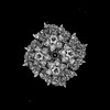
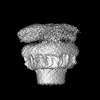
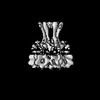
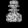
| Entry | Database: PDB / ID: 7wi4 | ||||||
|---|---|---|---|---|---|---|---|
| Title | Cryo-EM structure of E.Coli FtsH protease cytosolic domains | ||||||
 Components Components | ATP-dependent zinc metalloprotease FtsH | ||||||
 Keywords Keywords | HYDROLASE / complex / MEMBRANE PROTEIN | ||||||
| Function / homology |  Function and homology information Function and homology informationmembrane protein complex / plasma membrane protein complex / Hydrolases; Acting on peptide bonds (peptidases); Metalloendopeptidases / ATP-dependent peptidase activity / protein catabolic process / metalloendopeptidase activity / ATP hydrolysis activity / proteolysis / zinc ion binding / ATP binding / plasma membrane Similarity search - Function | ||||||
| Biological species |  | ||||||
| Method | ELECTRON MICROSCOPY / single particle reconstruction / cryo EM / Resolution: 3.4 Å | ||||||
 Authors Authors | Qiao, Z. / Gao, Y.G. | ||||||
| Funding support |  Singapore, 1items Singapore, 1items
| ||||||
 Citation Citation | Journal: Cell Rep / Year: 2022 Title: Cryo-EM structure of the entire FtsH-HflKC AAA protease complex. Authors: Zhu Qiao / Tatsuhiko Yokoyama / Xin-Fu Yan / Ing Tsyr Beh / Jian Shi / Sandip Basak / Yoshinori Akiyama / Yong-Gui Gao /  Abstract: The membrane-bound AAA protease FtsH is the key player controlling protein quality in bacteria. Two single-pass membrane proteins, HflK and HflC, interact with FtsH to modulate its proteolytic ...The membrane-bound AAA protease FtsH is the key player controlling protein quality in bacteria. Two single-pass membrane proteins, HflK and HflC, interact with FtsH to modulate its proteolytic activity. Here, we present structure of the entire FtsH-HflKC complex, comprising 12 copies of both HflK and HflC, all of which interact reciprocally to form a cage, as well as four FtsH hexamers with periplasmic domains and transmembrane helices enclosed inside the cage and cytoplasmic domains situated at the base of the cage. FtsH K61/D62/S63 in the β2-β3 loop in the periplasmic domain directly interact with HflK, contributing to complex formation. Pull-down and in vivo enzymatic activity assays validate the importance of the interacting interface for FtsH-HflKC complex formation. Structural comparison with the substrate-bound human m-AAA protease AFG3L2 offers implications for the HflKC cage in modulating substrate access to FtsH. Together, our findings provide a better understanding of FtsH-type AAA protease holoenzyme assembly and regulation. | ||||||
| History |
|
- Structure visualization
| Structure viewer | Molecule: MolmilJmol/JSmol |
|---|
- Downloads & links
-Download
| PDBx/mmCIF format | 7wi4.cif.gz | 435.3 KB | Display | PDBx/mmCIF format |
|---|---|---|---|---|
| PDB format | pdb7wi4.ent.gz | 353.3 KB | Display | PDB format |
| PDBx/mmJSON format | 7wi4.json.gz | Tree view | PDBx/mmJSON format | |
| Others |  Other downloads Other downloads |
-Validation report
| Arichive directory | https://data.pdbj.org/pub/pdb/validation_reports/wi/7wi4ftp://data.pdbj.org/pub/pdb/validation_reports/wi/7wi4 | HTTPS FTP |
|---|
-Related structure data
| Related structure data | 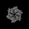 32521MC  7wi3C M: map data used to model this data C: citing same article ( |
|---|---|
| Similar structure data |
-Links
 PDBj
PDBj


- Assembly
| Deposited unit | 
|
|---|---|
| 1 |
|
-Components
| #1: Protein | Mass: 70795.055 Da / Num. of mol.: 6 / Mutation: E252Q, E415Q Source method: isolated from a genetically manipulated source Source: (gene. exp.) References: UniProt: P0AAI3, Hydrolases; Acting on peptide bonds (peptidases); Metalloendopeptidases #2: Chemical | ChemComp-ANP /   Mass: 506.196 Da / Num. of mol.: 6 / Source method: obtained synthetically / Formula: C10H17N6O12P3 / Comment: AMP-PNP, energy-carrying molecule analogue*YM Mass: 506.196 Da / Num. of mol.: 6 / Source method: obtained synthetically / Formula: C10H17N6O12P3 / Comment: AMP-PNP, energy-carrying molecule analogue*YM#3: Chemical | ChemComp-ZN /   Mass: 65.409 Da / Num. of mol.: 6 / Source method: obtained synthetically / Formula: Zn Mass: 65.409 Da / Num. of mol.: 6 / Source method: obtained synthetically / Formula: Zn#4: Chemical | ChemComp-MG /   Mass: 24.305 Da / Num. of mol.: 6 / Source method: obtained synthetically / Formula: Mg Mass: 24.305 Da / Num. of mol.: 6 / Source method: obtained synthetically / Formula: MgHas ligand of interest | N | |
|---|
-Experimental details
-Experiment
| Experiment | Method: ELECTRON MICROSCOPY |
|---|---|
| EM experiment | Aggregation state: PARTICLE / 3D reconstruction method: single particle reconstruction |
- Sample preparation
| Component | Name: E.Coli protein complex / Type: COMPLEX / Entity ID: #1 / Source: RECOMBINANT |
|---|---|
| Source (natural) | Organism: |
| Source (recombinant) | Organism: |
| Buffer solution | pH: 7.5 |
| Specimen | Conc.: 0.6 mg/ml / Embedding applied: NO / Shadowing applied: NO / Staining applied: NO / Vitrification applied: YES |
| Vitrification | Cryogen name: ETHANE |
- Electron microscopy imaging
| Experimental equipment |  Model: Titan Krios / Image courtesy: FEI Company |
|---|---|
| Microscopy | Model: FEI TITAN KRIOS |
| Electron gun | Electron source:  FIELD EMISSION GUN / Accelerating voltage: 300 kV / Illumination mode: OTHER FIELD EMISSION GUN / Accelerating voltage: 300 kV / Illumination mode: OTHER |
| Electron lens | Mode: BRIGHT FIELD / Nominal defocus max: 1500 nm / Nominal defocus min: 500 nm |
| Image recording | Electron dose: 60 e/Å2 / Film or detector model: GATAN K3 (6k x 4k) |
- Processing
| Software | Name: PHENIX / Version: 1.18.2_3874: / Classification: refinement | ||||||||||||||||||||||||
|---|---|---|---|---|---|---|---|---|---|---|---|---|---|---|---|---|---|---|---|---|---|---|---|---|---|
| CTF correction | Type: PHASE FLIPPING ONLY | ||||||||||||||||||||||||
| 3D reconstruction | Resolution: 3.4 Å / Resolution method: FSC 0.143 CUT-OFF / Num. of particles: 32359 / Symmetry type: POINT | ||||||||||||||||||||||||
| Refinement | Cross valid method: NONE Stereochemistry target values: GeoStd + Monomer Library + CDL v1.2 | ||||||||||||||||||||||||
| Displacement parameters | Biso mean: 80 Å2 | ||||||||||||||||||||||||
| Refine LS restraints |
|
