 Movie
Movie Controller

[English] 日本語
 Yorodumi
- PDB-7whs: Cryo-EM Structure of Leishmanial GDP-mannose pyrophosphorylase in... -
+ Open data

- Basic information
| Entry | Database: PDB / ID: 7whs | |||||||||
|---|---|---|---|---|---|---|---|---|---|---|
| Title | Cryo-EM Structure of Leishmanial GDP-mannose pyrophosphorylase in complex with GTP | |||||||||
 Components Components | Nucleotidyl transferase family protein | |||||||||
 Keywords Keywords | SUGAR BINDING PROTEIN / protozoan enzyme / Leishmania donovani | |||||||||
| Function / homology |  Function and homology information Function and homology informationmannose-1-phosphate guanylyltransferase / mannose-1-phosphate guanylyltransferase (GTP) activity / GDP-mannose biosynthetic process / GTP binding Similarity search - Function | |||||||||
| Biological species |  Leishmania donovani (eukaryote) Leishmania donovani (eukaryote) | |||||||||
| Method | ELECTRON MICROSCOPY / single particle reconstruction / cryo EM / Resolution: 3.1 Å | |||||||||
 Authors Authors | Xu, W. / Li, H. / Huang, C. | |||||||||
| Funding support |  China, 2items China, 2items
| |||||||||
 Citation Citation | Journal: Cell Discov / Year: 2022 Title: Structural insights into selective inhibition of leishmanial GDP-mannose pyrophosphorylase. Authors: Hang Li / Tuo Ji / Qi Sun / Yao Chen / Weiya Xu / Chengdong Huang / | |||||||||
| History |
|
- Structure visualization
| Structure viewer | Molecule: MolmilJmol/JSmol |
|---|
- Downloads & links
-Download
| PDBx/mmCIF format | 7whs.cif.gz | 375 KB | Display | PDBx/mmCIF format |
|---|---|---|---|---|
| PDB format | pdb7whs.ent.gz | 310 KB | Display | PDB format |
| PDBx/mmJSON format | 7whs.json.gz | Tree view | PDBx/mmJSON format | |
| Others |  Other downloads Other downloads |
-Validation report
| Arichive directory | https://data.pdbj.org/pub/pdb/validation_reports/wh/7whsftp://data.pdbj.org/pub/pdb/validation_reports/wh/7whs | HTTPS FTP |
|---|
-Related structure data
| Related structure data |  32510MC  7whrC  7whtC M: map data used to model this data C: citing same article ( |
|---|---|
| Similar structure data |


-Links
 PDBj
PDBj

- Assembly
| Deposited unit | 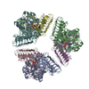
|
|---|---|
| 1 |
|
-Components
| #1: Protein | Mass: 41780.074 Da / Num. of mol.: 6 Source method: isolated from a genetically manipulated source Source: (gene. exp.) Leishmania donovani (eukaryote) / Gene: CGC21_17635 / Production host:  #2: Chemical | ChemComp-GTP /   Mass: 523.180 Da / Num. of mol.: 6 / Source method: obtained synthetically / Formula: C10H16N5O14P3 / Feature type: SUBJECT OF INVESTIGATION / Comment: GTP, energy-carrying molecule*YM Mass: 523.180 Da / Num. of mol.: 6 / Source method: obtained synthetically / Formula: C10H16N5O14P3 / Feature type: SUBJECT OF INVESTIGATION / Comment: GTP, energy-carrying molecule*YM#3: Chemical | ChemComp-MG /   Mass: 24.305 Da / Num. of mol.: 6 / Source method: obtained synthetically / Formula: Mg / Feature type: SUBJECT OF INVESTIGATION Mass: 24.305 Da / Num. of mol.: 6 / Source method: obtained synthetically / Formula: Mg / Feature type: SUBJECT OF INVESTIGATIONHas ligand of interest | Y | |
|---|
-Experimental details
-Experiment
| Experiment | Method: ELECTRON MICROSCOPY |
|---|---|
| EM experiment | Aggregation state: PARTICLE / 3D reconstruction method: single particle reconstruction |
- Sample preparation
| Component | Name: Ld GDP-MP / Type: CELL / Entity ID: #1 / Source: RECOMBINANT |
|---|---|
| Source (natural) | Organism: Leishmania donovani (eukaryote) |
| Source (recombinant) | Organism: |
| Buffer solution | pH: 7.5 |
| Specimen | Embedding applied: NO / Shadowing applied: NO / Staining applied: NO / Vitrification applied: YES |
| Vitrification | Cryogen name: ETHANE |
- Electron microscopy imaging
| Experimental equipment |  Model: Titan Krios / Image courtesy: FEI Company |
|---|---|
| Microscopy | Model: FEI TITAN KRIOS |
| Electron gun | Electron source:  FIELD EMISSION GUN / Accelerating voltage: 300 kV / Illumination mode: FLOOD BEAM FIELD EMISSION GUN / Accelerating voltage: 300 kV / Illumination mode: FLOOD BEAM |
| Electron lens | Mode: BRIGHT FIELD / Nominal defocus max: 2300 nm / Nominal defocus min: 1600 nm |
| Image recording | Electron dose: 51 e/Å2 / Film or detector model: GATAN K3 BIOQUANTUM (6k x 4k) |
- Processing
| Software | Name: PHENIX / Version: 1.19_4092: / Classification: refinement | ||||||||||||||||||||||||
|---|---|---|---|---|---|---|---|---|---|---|---|---|---|---|---|---|---|---|---|---|---|---|---|---|---|
| CTF correction | Type: NONE | ||||||||||||||||||||||||
| 3D reconstruction | Resolution: 3.1 Å / Resolution method: FSC 0.143 CUT-OFF / Num. of particles: 271350 / Symmetry type: POINT | ||||||||||||||||||||||||
| Refine LS restraints |
|
