 Movie
Movie Controller


+ Open data

- Basic information
| Entry | Database: PDB / ID: 7w8j | ||||||
|---|---|---|---|---|---|---|---|
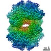
| Title | Dimethylformamidase, 2x(A2B2) | ||||||
 Components Components |
| ||||||
 Keywords Keywords | HYDROLASE / Amidohydrolase / mononuclear iron / AB polypeptide / tetramer | ||||||
| Function / homology |  Function and homology information Function and homology informationN,N-dimethylformamidase / N,N-dimethylformamidase activity / metal ion binding Similarity search - Function | ||||||
| Biological species |  Paracoccus sp. SSG05 (bacteria) Paracoccus sp. SSG05 (bacteria) | ||||||
| Method | ELECTRON MICROSCOPY / single particle reconstruction / cryo EM / Resolution: 2.5 Å | ||||||
 Authors Authors | Vinothkumar, K.R. / Subramanian, R. / Arya, C. / Ramanathan, G. | ||||||
| Funding support |  India, 1items India, 1items
| ||||||
 Citation Citation | Journal: To Be Published Title: Dimethylformamidase with a Unique Iron Center Authors: Arya, C. / Ramanathan, G. / Vinothkumar, K.R. / Subramanian, R. | ||||||
| History |
|
- Structure visualization
| Structure viewer | Molecule: MolmilJmol/JSmol |
|---|
- Downloads & links
-Download
| PDBx/mmCIF format | 7w8j.cif.gz | 638.4 KB | Display | PDBx/mmCIF format |
|---|---|---|---|---|
| PDB format | pdb7w8j.ent.gz | 518.5 KB | Display | PDB format |
| PDBx/mmJSON format | 7w8j.json.gz | Tree view | PDBx/mmJSON format | |
| Others |  Other downloads Other downloads |
-Validation report
| Arichive directory | https://data.pdbj.org/pub/pdb/validation_reports/w8/7w8jftp://data.pdbj.org/pub/pdb/validation_reports/w8/7w8j | HTTPS FTP |
|---|
-Related structure data
| Related structure data |  32357MC M: map data used to model this data C: citing same article ( |
|---|---|
| Similar structure data |
-Links
 PDBj
PDBj- Assembly
| Deposited unit | 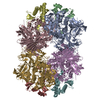
| |||||||||||||||||||||||||||||||||||||||||||||||||||||||||||||||||||||||||||||||||||||||||||||||||||||||||||||||||||||||||||||||||||||||||||||||||||||||||||||||||||||||||||||||||||||||||||||||||||||||||||||||||||||
|---|---|---|---|---|---|---|---|---|---|---|---|---|---|---|---|---|---|---|---|---|---|---|---|---|---|---|---|---|---|---|---|---|---|---|---|---|---|---|---|---|---|---|---|---|---|---|---|---|---|---|---|---|---|---|---|---|---|---|---|---|---|---|---|---|---|---|---|---|---|---|---|---|---|---|---|---|---|---|---|---|---|---|---|---|---|---|---|---|---|---|---|---|---|---|---|---|---|---|---|---|---|---|---|---|---|---|---|---|---|---|---|---|---|---|---|---|---|---|---|---|---|---|---|---|---|---|---|---|---|---|---|---|---|---|---|---|---|---|---|---|---|---|---|---|---|---|---|---|---|---|---|---|---|---|---|---|---|---|---|---|---|---|---|---|---|---|---|---|---|---|---|---|---|---|---|---|---|---|---|---|---|---|---|---|---|---|---|---|---|---|---|---|---|---|---|---|---|---|---|---|---|---|---|---|---|---|---|---|---|---|---|---|---|---|
| 1 |
| |||||||||||||||||||||||||||||||||||||||||||||||||||||||||||||||||||||||||||||||||||||||||||||||||||||||||||||||||||||||||||||||||||||||||||||||||||||||||||||||||||||||||||||||||||||||||||||||||||||||||||||||||||||
| Noncrystallographic symmetry (NCS) | NCS domain:
NCS domain segments: Component-ID: _ / Refine code: _
|