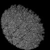
 Movie
Movie Controller


+ Open data

- Basic information
| Entry | Database: PDB / ID: 7vrp | |||||||||
|---|---|---|---|---|---|---|---|---|---|---|
| Title | Structure of infectious bursal disease virus Gx strain | |||||||||
 Components Components | Structural polyprotein | |||||||||
 Keywords Keywords | VIRUS / IBDV / icosahedral / very virulent | |||||||||
| Function / homology |  Function and homology information Function and homology informationHydrolases; Acting on peptide bonds (peptidases); Serine endopeptidases / serine-type peptidase activity / viral capsid / host cell cytoplasm / structural molecule activity / proteolysis / metal ion binding Similarity search - Function | |||||||||
| Biological species |  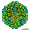 Avian infectious bursal disease virus (Gumboro virus) Avian infectious bursal disease virus (Gumboro virus) | |||||||||
| Method | ELECTRON MICROSCOPY / single particle reconstruction / cryo EM / Resolution: 3.8 Å | |||||||||
 Authors Authors | Bao, K.Y. / Zhu, P. | |||||||||
| Funding support |  China, 2items China, 2items
| |||||||||
 Citation Citation | Journal: Sci Bull (Beijing) / Year: 2022 Title: Cryo-EM structures of infectious bursal disease viruses with different virulences provide insights into their assembly and invasion Authors: Bao, K. / Qi, X. / Lia, Y. / Gong, M. / Wang, M. / Zhu, P. | |||||||||
| History |
|
- Structure visualization
| Structure viewer | Molecule: MolmilJmol/JSmol |
|---|
- Downloads & links
-Download
| PDBx/mmCIF format | 7vrp.cif.gz | 869.6 KB | Display | PDBx/mmCIF format |
|---|---|---|---|---|
| PDB format | pdb7vrp.ent.gz | 741.3 KB | Display | PDB format |
| PDBx/mmJSON format | 7vrp.json.gz | Tree view | PDBx/mmJSON format | |
| Others |  Other downloads Other downloads |
-Validation report
| Summary document | 7vrp_validation.pdf.gz | 1.2 MB | Display | wwPDB validaton report |
|---|---|---|---|---|
| Full document | 7vrp_full_validation.pdf.gz | 1.2 MB | Display | |
| Data in XML | 7vrp_validation.xml.gz | 131.5 KB | Display | |
| Data in CIF | 7vrp_validation.cif.gz | 202.9 KB | Display | |
| Arichive directory | https://data.pdbj.org/pub/pdb/validation_reports/vr/7vrpftp://data.pdbj.org/pub/pdb/validation_reports/vr/7vrp | HTTPS FTP |
-Related structure data
| Related structure data |  32102MC 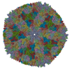 7vrnC M: map data used to model this data C: citing same article ( |
|---|---|
| Similar structure data |
-Links
 PDBj
PDBj- Assembly
| Deposited unit | 
|
|---|---|
| 1 | x 60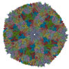
|
| 2 |
|
| 3 | x 5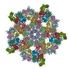
|
| 4 | x 6
|
| 5 | 
|
| Symmetry | Point symmetry: (Schoenflies symbol: I (icosahedral)) |
-Components
| #1: Protein | Mass: 47028.871 Da / Num. of mol.: 13 / Source method: isolated from a natural source Source: (natural) Avian infectious bursal disease virus (Gumboro virus)References: UniProt: Q6SZ77, Hydrolases; Acting on peptide bonds (peptidases); Serine endopeptidases |
|---|
-Experimental details
-Experiment
| Experiment | Method: ELECTRON MICROSCOPY |
|---|---|
| EM experiment | Aggregation state: PARTICLE / 3D reconstruction method: single particle reconstruction |
- Sample preparation
| Component | Name: Infectious bursal disease virus / Type: VIRUS / Entity ID: all / Source: NATURAL |
|---|---|
| Source (natural) | Organism: Infectious bursal disease virus (Gumboro virus) |
| Details of virus | Empty: NO / Enveloped: NO / Isolate: STRAIN / Type: VIRION |
| Natural host | Organism: Gallus gallus |
| Buffer solution | pH: 7.2 |
| Specimen | Embedding applied: NO / Shadowing applied: NO / Staining applied: NO / Vitrification applied: YES |
| Vitrification | Cryogen name: ETHANE |
- Electron microscopy imaging
| Experimental equipment |  Model: Titan Krios / Image courtesy: FEI Company |
|---|---|
| Microscopy | Model: FEI TITAN KRIOS |
| Electron gun | Electron source:  FIELD EMISSION GUN / Accelerating voltage: 300 kV / Illumination mode: FLOOD BEAM FIELD EMISSION GUN / Accelerating voltage: 300 kV / Illumination mode: FLOOD BEAM |
| Electron lens | Mode: BRIGHT FIELD |
| Image recording | Electron dose: 47.5 e/Å2 / Film or detector model: GATAN K2 SUMMIT (4k x 4k) |
- Processing
| CTF correction | Type: PHASE FLIPPING AND AMPLITUDE CORRECTION |
|---|---|
| 3D reconstruction | Resolution: 3.8 Å / Resolution method: FSC 0.143 CUT-OFF / Num. of particles: 3524 / Symmetry type: POINT |
