Movie
Movie Controller

[English] 日本語
 Yorodumi
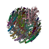
- PDB-7vrj: STRUCTURE OF PHOTOSYNTHETIC LH1-RC SUPER-COMPLEX OF Allochromatiu... -
+ Open data

- Basic information
| Entry | Database: PDB / ID: 7vrj | |||||||||||||||||||||
|---|---|---|---|---|---|---|---|---|---|---|---|---|---|---|---|---|---|---|---|---|---|---|
| Title | STRUCTURE OF PHOTOSYNTHETIC LH1-RC SUPER-COMPLEX OF Allochromatium tepidum | |||||||||||||||||||||
 Components Components |
| |||||||||||||||||||||
 Keywords Keywords | PHOTOSYNTHESIS / LH1-RC COMPLEX / PURPLE BACTERIA | |||||||||||||||||||||
| Function / homology |  Function and homology information Function and homology informationPhotosynthetic Reaction Center; Chain H, domain 2 / Photosynthetic Reaction Center, subunit H, domain 2 / Alpha-Beta Complex / Alpha Beta Similarity search - Domain/homology | |||||||||||||||||||||
| Biological species |  Allochromatium tepidum (bacteria) Allochromatium tepidum (bacteria) | |||||||||||||||||||||
| Method | ELECTRON MICROSCOPY / single particle reconstruction / cryo EM / Resolution: 2.81 Å | |||||||||||||||||||||
 Authors Authors | Tani, K. / Kobayashi, K. / Hosogi, N. / Ji, X.-C. / Nagashima, S. / Nagashima, K.V.P. / Tsukatani, Y. / Kanno, R. / Hall, M. / Yu, L.-J. ...Tani, K. / Kobayashi, K. / Hosogi, N. / Ji, X.-C. / Nagashima, S. / Nagashima, K.V.P. / Tsukatani, Y. / Kanno, R. / Hall, M. / Yu, L.-J. / Ishikawa, I. / Okura, Y. / Madigan, M.T. / Mizoguchi, A. / Humbel, B.M. / Kimura, Y. / Wang-Otomo, Z.-Y. | |||||||||||||||||||||
| Funding support |  Japan, 6items Japan, 6items
| |||||||||||||||||||||
 Citation Citation | Journal: J Biol Chem / Year: 2022 Title: A Ca-binding motif underlies the unusual properties of certain photosynthetic bacterial core light-harvesting complexes. Authors: Kazutoshi Tani / Kazumi Kobayashi / Naoki Hosogi / Xuan-Cheng Ji / Sakiko Nagashima / Kenji V P Nagashima / Airi Izumida / Kazuhito Inoue / Yusuke Tsukatani / Ryo Kanno / Malgorzata Hall / ...Authors: Kazutoshi Tani / Kazumi Kobayashi / Naoki Hosogi / Xuan-Cheng Ji / Sakiko Nagashima / Kenji V P Nagashima / Airi Izumida / Kazuhito Inoue / Yusuke Tsukatani / Ryo Kanno / Malgorzata Hall / Long-Jiang Yu / Isamu Ishikawa / Yoshihiro Okura / Michael T Madigan / Akira Mizoguchi / Bruno M Humbel / Yukihiro Kimura / Zheng-Yu Wang-Otomo /   Abstract: The mildly thermophilic purple phototrophic bacterium Allochromatium tepidum provides a unique model for investigating various intermediate phenotypes observed between those of thermophilic and ...The mildly thermophilic purple phototrophic bacterium Allochromatium tepidum provides a unique model for investigating various intermediate phenotypes observed between those of thermophilic and mesophilic counterparts. The core light-harvesting (LH1) complex from A. tepidum exhibits an absorption maximum at 890 nm and mildly enhanced thermostability, both of which are Ca-dependent. However, it is unknown what structural determinants might contribute to these properties. Here, we present a cryo-EM structure of the reaction center-associated LH1 complex at 2.81 Å resolution, in which we identify multiple pigment-binding α- and β-polypeptides within an LH1 ring. Of the 16 α-polypeptides, we show that six (α1) bind Ca along with β1- or β3-polypeptides to form the Ca-binding sites. This structure differs from that of fully Ca-bound LH1 from Thermochromatium tepidum, enabling determination of the minimum structural requirements for Ca-binding. We also identified three amino acids (Trp44, Asp47, and Ile49) in the C-terminal region of the A. tepidum α1-polypeptide that ligate each Ca ion, forming a Ca-binding WxxDxI motif that is conserved in all Ca-bound LH1 α-polypeptides from other species with reported structures. The partial Ca-bound structure further explains the unusual phenotypic properties observed for this bacterium in terms of its Ca-requirements for thermostability, spectroscopy, and phototrophic growth, and supports the hypothesis that A. tepidum may represent a "transitional" species between mesophilic and thermophilic purple sulfur bacteria. The characteristic arrangement of multiple αβ-polypeptides also suggests a mechanism of molecular recognition in the expression and/or assembly of the LH1 complex that could be regulated through interactions with reaction center subunits. | |||||||||||||||||||||
| History |
|
- Structure visualization
| Structure viewer | Molecule: MolmilJmol/JSmol |
|---|
- Downloads & links
-Download
| PDBx/mmCIF format | 7vrj.cif.gz | 567.7 KB | Display | PDBx/mmCIF format |
|---|---|---|---|---|
| PDB format | pdb7vrj.ent.gz | 511.9 KB | Display | PDB format |
| PDBx/mmJSON format | 7vrj.json.gz | Tree view | PDBx/mmJSON format | |
| Others |  Other downloads Other downloads |
-Validation report
| Summary document | 7vrj_validation.pdf.gz | 5 MB | Display | wwPDB validaton report |
|---|---|---|---|---|
| Full document | 7vrj_full_validation.pdf.gz | 5.2 MB | Display | |
| Data in XML | 7vrj_validation.xml.gz | 133.1 KB | Display | |
| Data in CIF | 7vrj_validation.cif.gz | 167 KB | Display | |
| Arichive directory | https://data.pdbj.org/pub/pdb/validation_reports/vr/7vrjftp://data.pdbj.org/pub/pdb/validation_reports/vr/7vrj | HTTPS FTP |
-Related structure data
| Related structure data |  32100MC M: map data used to model this data C: citing same article ( |
|---|---|
| Similar structure data |
-Links
 PDBj
PDBj


- Assembly
| Deposited unit | 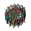
|
|---|---|
| 1 |
|
-Components
-Photosynthetic reaction center ... , 4 types, 4 molecules CLMH
| #1: Protein | Mass: 43659.016 Da / Num. of mol.: 1 / Source method: isolated from a natural source / Source: (natural) Allochromatium tepidum (bacteria) |
|---|---|
| #2: Protein | Mass: 31098.203 Da / Num. of mol.: 1 / Source method: isolated from a natural source / Source: (natural) Allochromatium tepidum (bacteria) |
| #3: Protein | Mass: 36292.117 Da / Num. of mol.: 1 / Source method: isolated from a natural source / Source: (natural) Allochromatium tepidum (bacteria) |
| #4: Protein | Mass: 28196.340 Da / Num. of mol.: 1 / Source method: isolated from a natural source / Source: (natural) Allochromatium tepidum (bacteria) |
-Light-harvesting protein LH1 ... , 5 types, 32 molecules AIKOQ1579BJNPR24680DFSUWYEGTVXZ3
| #5: Protein/peptide | Mass: 5136.131 Da / Num. of mol.: 9 / Source method: isolated from a natural source / Source: (natural) Allochromatium tepidum (bacteria)#6: Protein/peptide | Mass: 5269.103 Da / Num. of mol.: 10 / Source method: isolated from a natural source / Source: (natural) Allochromatium tepidum (bacteria)#7: Protein | Mass: 7330.739 Da / Num. of mol.: 6 / Source method: isolated from a natural source / Source: (natural) Allochromatium tepidum (bacteria)#8: Protein/peptide | Mass: 5504.446 Da / Num. of mol.: 6 / Source method: isolated from a natural source / Source: (natural) Allochromatium tepidum (bacteria)#9: Protein | | Mass: 7323.753 Da / Num. of mol.: 1 / Source method: isolated from a natural source / Source: (natural) Allochromatium tepidum (bacteria) |
|---|
-Sugars , 1 types, 18 molecules 
| #22: Sugar | ChemComp-LMT /  Type: D-saccharide / Mass: 510.615 Da / Num. of mol.: 18 / Source method: obtained synthetically / Formula: C24H46O11 / Comment: detergent*YM Type: D-saccharide / Mass: 510.615 Da / Num. of mol.: 18 / Source method: obtained synthetically / Formula: C24H46O11 / Comment: detergent*YM |
|---|
-Non-polymers , 15 types, 144 molecules 

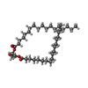


| #10: Chemical | ChemComp-HEC /  Mass: 618.503 Da / Num. of mol.: 4 / Source method: obtained synthetically / Formula: C34H34FeN4O4 Mass: 618.503 Da / Num. of mol.: 4 / Source method: obtained synthetically / Formula: C34H34FeN4O4#11: Chemical | ChemComp-MG / |  Mass: 24.305 Da / Num. of mol.: 1 / Source method: obtained synthetically / Formula: Mg Mass: 24.305 Da / Num. of mol.: 1 / Source method: obtained synthetically / Formula: Mg#12: Chemical | ChemComp-DGA / |  Mass: 625.018 Da / Num. of mol.: 1 / Source method: obtained synthetically / Formula: C39H76O5 Mass: 625.018 Da / Num. of mol.: 1 / Source method: obtained synthetically / Formula: C39H76O5#13: Chemical | ChemComp-PLM / |  Mass: 256.424 Da / Num. of mol.: 1 / Source method: obtained synthetically / Formula: C16H32O2 Mass: 256.424 Da / Num. of mol.: 1 / Source method: obtained synthetically / Formula: C16H32O2#14: Chemical | ChemComp-PGV / (  Mass: 749.007 Da / Num. of mol.: 12 / Source method: obtained synthetically / Formula: C40H77O10P / Comment: phospholipid*YM Mass: 749.007 Da / Num. of mol.: 12 / Source method: obtained synthetically / Formula: C40H77O10P / Comment: phospholipid*YM#15: Chemical | ChemComp-BCL /  Mass: 911.504 Da / Num. of mol.: 36 / Source method: obtained synthetically / Formula: C55H74MgN4O6 / Feature type: SUBJECT OF INVESTIGATION Mass: 911.504 Da / Num. of mol.: 36 / Source method: obtained synthetically / Formula: C55H74MgN4O6 / Feature type: SUBJECT OF INVESTIGATION#16: Chemical |  Mass: 889.215 Da / Num. of mol.: 2 / Source method: obtained synthetically / Formula: C55H76N4O6 Mass: 889.215 Da / Num. of mol.: 2 / Source method: obtained synthetically / Formula: C55H76N4O6#17: Chemical |  Mass: 727.109 Da / Num. of mol.: 3 / Source method: obtained synthetically / Formula: C49H74O4 Mass: 727.109 Da / Num. of mol.: 3 / Source method: obtained synthetically / Formula: C49H74O4#18: Chemical | ChemComp-CDL /  Mass: 1464.043 Da / Num. of mol.: 8 / Source method: obtained synthetically / Formula: C81H156O17P2 / Comment: phospholipid*YM Mass: 1464.043 Da / Num. of mol.: 8 / Source method: obtained synthetically / Formula: C81H156O17P2 / Comment: phospholipid*YM#19: Chemical | ChemComp-FE / |  Mass: 55.845 Da / Num. of mol.: 1 / Source method: obtained synthetically / Formula: Fe Mass: 55.845 Da / Num. of mol.: 1 / Source method: obtained synthetically / Formula: Fe#20: Chemical |  Mass: 717.116 Da / Num. of mol.: 2 / Source method: obtained synthetically / Formula: C51H72O2 Mass: 717.116 Da / Num. of mol.: 2 / Source method: obtained synthetically / Formula: C51H72O2#21: Chemical | ChemComp-CRT /  Mass: 596.925 Da / Num. of mol.: 17 / Source method: obtained synthetically / Formula: C42H60O2 Mass: 596.925 Da / Num. of mol.: 17 / Source method: obtained synthetically / Formula: C42H60O2#23: Chemical | ChemComp-CA /  Mass: 40.078 Da / Num. of mol.: 6 / Source method: obtained synthetically / Formula: Ca Mass: 40.078 Da / Num. of mol.: 6 / Source method: obtained synthetically / Formula: Ca#24: Chemical |  Mass: 229.402 Da / Num. of mol.: 2 / Source method: obtained synthetically / Formula: C14H31NO / Comment: LDAO, detergent*YM Mass: 229.402 Da / Num. of mol.: 2 / Source method: obtained synthetically / Formula: C14H31NO / Comment: LDAO, detergent*YM#25: Water | ChemComp-HOH / | Mass: 18.015 Da / Num. of mol.: 48 / Source method: isolated from a natural source / Formula: H2O |
|---|
-Details
| Has ligand of interest | Y |
|---|
-Experimental details
-Experiment
| Experiment | Method: ELECTRON MICROSCOPY |
|---|---|
| EM experiment | Aggregation state: PARTICLE / 3D reconstruction method: single particle reconstruction |
- Sample preparation
| Component | Name: Photosynthetic LH1-RC complex from the purple phototrophic bacterium Allochromatium tepidum Type: COMPLEX / Entity ID: #1-#9 / Source: NATURAL |
|---|---|
| Molecular weight | Units: MEGADALTONS / Experimental value: NO |
| Source (natural) | Organism: Allochromatium tepidum (bacteria) |
| Buffer solution | pH: 7.5 |
| Specimen | Conc.: 3 mg/ml / Embedding applied: NO / Shadowing applied: NO / Staining applied: NO / Vitrification applied: YES / Details: This sample was monodisperse. |
| Vitrification | Instrument: LEICA EM GP / Cryogen name: ETHANE / Humidity: 90 % / Chamber temperature: 277 K |
- Electron microscopy imaging
| Microscopy | Model: JEOL CRYO ARM 300 |
|---|---|
| Electron gun | Electron source:  FIELD EMISSION GUN / Accelerating voltage: 300 kV / Illumination mode: FLOOD BEAM FIELD EMISSION GUN / Accelerating voltage: 300 kV / Illumination mode: FLOOD BEAM |
| Electron lens | Mode: BRIGHT FIELD / Alignment procedure: BASIC |
| Specimen holder | Cryogen: NITROGEN / Specimen holder model: JEOL |
| Image recording | Average exposure time: 1.5 sec. / Electron dose: 40 e/Å2 / Detector mode: COUNTING / Film or detector model: GATAN K3 (6k x 4k) |
- Processing
| Software | Name: PHENIX / Version: 1.19.2_4158: / Classification: refinement | ||||||||||||||||||||||||||||||||||||
|---|---|---|---|---|---|---|---|---|---|---|---|---|---|---|---|---|---|---|---|---|---|---|---|---|---|---|---|---|---|---|---|---|---|---|---|---|---|
| EM software |
| ||||||||||||||||||||||||||||||||||||
| CTF correction | Type: PHASE FLIPPING ONLY | ||||||||||||||||||||||||||||||||||||
| Particle selection | Num. of particles selected: 309706 | ||||||||||||||||||||||||||||||||||||
| Symmetry | Point symmetry: C1 (asymmetric) | ||||||||||||||||||||||||||||||||||||
| 3D reconstruction | Resolution: 2.81 Å / Resolution method: FSC 0.143 CUT-OFF / Num. of particles: 156992 / Symmetry type: POINT | ||||||||||||||||||||||||||||||||||||
| Atomic model building | B value: 62 / Protocol: RIGID BODY FIT / Space: REAL / Target criteria: Correlation coefficient | ||||||||||||||||||||||||||||||||||||
| Atomic model building | PDB-ID: 5Y5S | ||||||||||||||||||||||||||||||||||||
| Refine LS restraints |
|