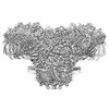
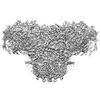
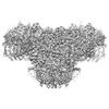
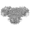
- PDB-7vmm: Structure of recombinant RyR2 (EGTA dataset, class 1, closed state) -
+
データを開く
IDまたはキーワード:
読み込み中...
-
基本情報
登録情報
データベース: PDB / ID: 7vmm
タイトル
Structure of recombinant RyR2 (EGTA dataset, class 1, closed state)
要素
Peptidyl-prolyl cis-trans isomerase FKBP1B
Ryanodine receptor 2
キーワード
MEMBRANE PROTEIN / CALCIUM / CALCIUM CHANNEL / CALCIUM TRANSPORT / ION TRANSPORT / IONIC CHANNEL / METAL TRANSPORT / ER/SR MEMBRANE / RYANODINE RECEPTOR / RYANODINE / RECEPTOR / WILD TYPE
機能・相同性
機能・相同性情報
manganese ion transmembrane transport / suramin binding / establishment of protein localization to endoplasmic reticulum / type B pancreatic cell apoptotic process / Purkinje myocyte to ventricular cardiac muscle cell signaling / regulation of SA node cell action potential / regulation of atrial cardiac muscle cell action potential / left ventricular cardiac muscle tissue morphogenesis / organic cyclic compound binding / regulation of AV node cell action potential ...manganese ion transmembrane transport / suramin binding / establishment of protein localization to endoplasmic reticulum / type B pancreatic cell apoptotic process / Purkinje myocyte to ventricular cardiac muscle cell signaling / regulation of SA node cell action potential / regulation of atrial cardiac muscle cell action potential / left ventricular cardiac muscle tissue morphogenesis / organic cyclic compound binding / regulation of AV node cell action potential / calcium-induced calcium release activity / sarcoplasmic reticulum calcium ion transport / Stimuli-sensing channels / Ion homeostasis / ventricular cardiac muscle cell action potential / regulation of ventricular cardiac muscle cell action potential / positive regulation of sequestering of calcium ion / cyclic nucleotide binding / embryonic heart tube morphogenesis / cardiac muscle hypertrophy / negative regulation of insulin secretion involved in cellular response to glucose stimulus / negative regulation of release of sequestered calcium ion into cytosol / ryanodine-sensitive calcium-release channel activity / neuronal action potential propagation / insulin secretion involved in cellular response to glucose stimulus / release of sequestered calcium ion into cytosol by sarcoplasmic reticulum / response to muscle activity / calcium ion transport into cytosol / calcium ion transmembrane import into cytosol / response to caffeine / cell communication by electrical coupling involved in cardiac conduction / A band / response to redox state / protein maturation by protein folding / 'de novo' protein folding / negative regulation of heart rate / negative regulation of phosphoprotein phosphatase activity / positive regulation of heart rate / FK506 binding / negative regulation of cytosolic calcium ion concentration / positive regulation of axon regeneration / cellular response to caffeine / protein kinase A regulatory subunit binding / intracellularly gated calcium channel activity / protein kinase A catalytic subunit binding / positive regulation of the force of heart contraction / response to magnesium ion / : / detection of calcium ion / smooth muscle contraction / smooth endoplasmic reticulum / negative regulation of ryanodine-sensitive calcium-release channel activity / response to vitamin E / calcium channel inhibitor activity / regulation of cardiac muscle contraction by regulation of the release of sequestered calcium ion / protein peptidyl-prolyl isomerization / T cell proliferation / striated muscle contraction / regulation of release of sequestered calcium ion into cytosol by sarcoplasmic reticulum / release of sequestered calcium ion into cytosol / Ion homeostasis / regulation of ryanodine-sensitive calcium-release channel activity / monoatomic ion transmembrane transport / extrinsic component of cytoplasmic side of plasma membrane / sarcoplasmic reticulum membrane / calcium channel complex / cellular response to epinephrine stimulus / regulation of cytosolic calcium ion concentration / response to muscle stretch / regulation of heart rate / sarcomere / peptidylprolyl isomerase / sarcoplasmic reticulum / peptidyl-prolyl cis-trans isomerase activity / establishment of localization in cell / calcium-mediated signaling / calcium ion transmembrane transport / calcium channel activity / response to hydrogen peroxide / Stimuli-sensing channels / sarcolemma / Z disc / intracellular calcium ion homeostasis / response to calcium ion / calcium ion transport / : / nuclear envelope / positive regulation of cytosolic calcium ion concentration / protein refolding / scaffold protein binding / transmembrane transporter binding / calmodulin binding / response to hypoxia / signaling receptor binding / calcium ion binding / protein kinase binding / enzyme binding / protein-containing complex / identical protein binding / membrane 類似検索 - 分子機能
Japan Agency for Medical Research and Development (AMED)
JP21am0101080
日本
Japan Agency for Medical Research and Development (AMED)
19ek0109202
日本
引用
ジャーナル: Nat Commun / 年: 2022 タイトル: Molecular basis for gating of cardiac ryanodine receptor explains the mechanisms for gain- and loss-of function mutations. 著者: Takuya Kobayashi / Akihisa Tsutsumi / Nagomi Kurebayashi / Kei Saito / Masami Kodama / Takashi Sakurai / Masahide Kikkawa / Takashi Murayama / Haruo Ogawa / 要旨: Cardiac ryanodine receptor (RyR2) is a large Ca release channel in the sarcoplasmic reticulum and indispensable for excitation-contraction coupling in the heart. RyR2 is activated by Ca and RyR2 ...Cardiac ryanodine receptor (RyR2) is a large Ca release channel in the sarcoplasmic reticulum and indispensable for excitation-contraction coupling in the heart. RyR2 is activated by Ca and RyR2 mutations are implicated in severe arrhythmogenic diseases. Yet, the structural basis underlying channel opening and how mutations affect the channel remains unknown. Here, we address the gating mechanism of RyR2 by combining high-resolution structures determined by cryo-electron microscopy with quantitative functional analysis of channels carrying various mutations in specific residues. We demonstrated two fundamental mechanisms for channel gating: interactions close to the channel pore stabilize the channel to prevent hyperactivity and a series of interactions in the surrounding regions is necessary for channel opening upon Ca binding. Mutations at the residues involved in the former and the latter mechanisms cause gain-of-function and loss-of-function, respectively. Our results reveal gating mechanisms of the RyR2 channel and alterations by pathogenic mutations at the atomic level.
履歴
登録
2021年10月9日
登録サイト: PDBJ / 処理サイト: PDBJ
改定 1.0
2022年8月10日
Provider: repository / タイプ: Initial release
改定 1.1
2024年6月19日
Group: Data collection / カテゴリ: chem_comp_atom / chem_comp_bond
 ムービー
ムービー コントローラー


 データを開く
 基本情報
基本情報 要素
要素 キーワード
キーワード 機能・相同性情報
 Homo sapiens (ヒト)
 データ登録者
 日本, 6件
  引用
引用 構造の表示
構造の表示 ダウンロードとリンク
 その他のダウンロード
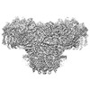


 PDBj
PDBj


 集合体
集合体


 分子量: 65.409 Da / 分子数: 4 / 由来タイプ: 合成 / 式: Zn / タイプ: SUBJECT OF INVESTIGATION
 試料調製
試料調製 電子顕微鏡撮影
 FIELD EMISSION GUN / 加速電圧: 300 kV / 照射モード: FLOOD BEAM
 解析
解析