 Movie
Movie Controller

[English] 日本語
 Yorodumi
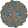
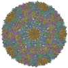
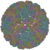
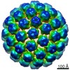
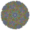
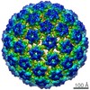
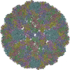
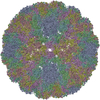
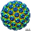
- PDB-7vi9: Cryo-EM structure of bacteriophage lambda procapsid at 5.03 Angstrom -
+ Open data

- Basic information
| Entry | Database: PDB / ID: 7vi9 | |||||||||||||||||||||||||||||||||||||||||||||
|---|---|---|---|---|---|---|---|---|---|---|---|---|---|---|---|---|---|---|---|---|---|---|---|---|---|---|---|---|---|---|---|---|---|---|---|---|---|---|---|---|---|---|---|---|---|---|
| Title | Cryo-EM structure of bacteriophage lambda procapsid at 5.03 Angstrom | |||||||||||||||||||||||||||||||||||||||||||||
 Components Components | Major capsid protein | |||||||||||||||||||||||||||||||||||||||||||||
 Keywords Keywords | VIRUS / bacteriophage lambda / capsid / procapsid / capsid maturation / virus structure / cryo-EM / auxiliary protein / conformational expansion / cementing protein / DNA packaging | |||||||||||||||||||||||||||||||||||||||||||||
| Function / homology | Major capsid protein GpE / Phage major capsid protein E / T=7 icosahedral viral capsid / viral capsid / host cell cytoplasm / Major capsid protein Function and homology information Function and homology information | |||||||||||||||||||||||||||||||||||||||||||||
| Biological species |  Escherichia phage lambda (virus) Escherichia phage lambda (virus) | |||||||||||||||||||||||||||||||||||||||||||||
| Method | ELECTRON MICROSCOPY / single particle reconstruction / cryo EM / Resolution: 5.03 Å | |||||||||||||||||||||||||||||||||||||||||||||
 Authors Authors | Wang, J.W. | |||||||||||||||||||||||||||||||||||||||||||||
| Funding support |  China, 2items China, 2items
| |||||||||||||||||||||||||||||||||||||||||||||
 Citation Citation | Journal: Structure / Year: 2022 Title: Structural basis of bacteriophage lambda capsid maturation. Authors: Chang Wang / Jianwei Zeng / Jiawei Wang / Abstract: Bacteriophage lambda is an excellent model system for studying capsid assembly of double-stranded DNA (dsDNA) bacteriophages, some dsDNA archaeal viruses, and herpesviruses. HK97 fold coat proteins ...Bacteriophage lambda is an excellent model system for studying capsid assembly of double-stranded DNA (dsDNA) bacteriophages, some dsDNA archaeal viruses, and herpesviruses. HK97 fold coat proteins initially assemble into a precursor capsid (procapsid) and subsequent genome packaging triggers morphological expansion of the shell. An auxiliary protein is required to stabilize the expanded capsid structure. To investigate the capsid maturation mechanism, we determined the cryo-electron microscopy structures of the bacteriophage lambda procapsid and mature capsid at 3.88 Å and 3.76 Å resolution, respectively. Besides primarily rigid body movements of common features of the major capsid protein gpE, large-scale structural rearrangements of other domains occur simultaneously. Assembly of intercapsomers within the procapsid is facilitated by layer-stacking effects at 3-fold vertices. Upon conformational expansion of the capsid shell, the missing top layer is fulfilled by cementing the gpD protein against the internal pressure of DNA packaging. Our structures illuminate the assembly mechanisms of dsDNA viruses. | |||||||||||||||||||||||||||||||||||||||||||||
| History |
|
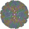
- Structure visualization
| Movie |
Movie viewer |
|---|---|
| Structure viewer | Molecule: MolmilJmol/JSmol |
- Downloads & links
-Download
| PDBx/mmCIF format | 7vi9.cif.gz | 403.1 KB | Display | PDBx/mmCIF format |
|---|---|---|---|---|
| PDB format | pdb7vi9.ent.gz | 337.8 KB | Display | PDB format |
| PDBx/mmJSON format | 7vi9.json.gz | Tree view | PDBx/mmJSON format | |
| Others |  Other downloads Other downloads |
-Validation report
| Arichive directory | https://data.pdbj.org/pub/pdb/validation_reports/vi/7vi9ftp://data.pdbj.org/pub/pdb/validation_reports/vi/7vi9 | HTTPS FTP |
|---|
-Related structure data
| Related structure data |  32004MC  7viaC  7viiC  7vikC M: map data used to model this data C: citing same article ( |
|---|---|
| Similar structure data |


-Links
 PDBj
PDBj
- Assembly
| Deposited unit | 
|
|---|---|
| 1 | x 60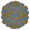
|
| 2 |
|
| 3 | x 5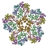
|
| 4 | x 6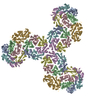
|
| 5 | 
|
| Symmetry | Point symmetry: (Schoenflies symbol: I (icosahedral)) |
-Components
| #1: Protein | Mass: 38229.160 Da / Num. of mol.: 7 Source method: isolated from a genetically manipulated source Source: (gene. exp.) Escherichia phage lambda (virus) / Gene: E, lambdap08 / Production host:  Has protein modification | N | |
|---|
-Experimental details
-Experiment
| Experiment | Method: ELECTRON MICROSCOPY |
|---|---|
| EM experiment | Aggregation state: PARTICLE / 3D reconstruction method: single particle reconstruction |
- Sample preparation
| Component | Name: Escherichia virus Lambda / Type: VIRUS / Entity ID: all / Source: NATURAL |
|---|---|
| Source (natural) | Organism: Escherichia virus Lambda |
| Details of virus | Empty: YES / Enveloped: NO / Isolate: OTHER / Type: VIRION |
| Buffer solution | pH: 7.4 |
| Specimen | Embedding applied: NO / Shadowing applied: NO / Staining applied: NO / Vitrification applied: YES |
| Vitrification | Cryogen name: NITROGEN |
- Electron microscopy imaging
| Experimental equipment |  Model: Titan Krios / Image courtesy: FEI Company |
|---|---|
| Microscopy | Model: FEI TITAN KRIOS |
| Electron gun | Electron source:  FIELD EMISSION GUN / Accelerating voltage: 300 kV / Illumination mode: OTHER FIELD EMISSION GUN / Accelerating voltage: 300 kV / Illumination mode: OTHER |
| Electron lens | Mode: BRIGHT FIELD |
| Image recording | Electron dose: 50 e/Å2 / Film or detector model: GATAN K3 (6k x 4k) |
- Processing
| Software | Name: PHENIX / Version: 1.19.2_4158: / Classification: refinement | ||||||||||||||||||||||||
|---|---|---|---|---|---|---|---|---|---|---|---|---|---|---|---|---|---|---|---|---|---|---|---|---|---|
| EM software | Name: PHENIX / Category: model refinement | ||||||||||||||||||||||||
| CTF correction | Type: NONE | ||||||||||||||||||||||||
| 3D reconstruction | Resolution: 5.03 Å / Resolution method: FSC 0.143 CUT-OFF / Num. of particles: 174346 / Symmetry type: POINT | ||||||||||||||||||||||||
| Atomic model building | Protocol: OTHER | ||||||||||||||||||||||||
| Refine LS restraints |
|
