 Movie
Movie Controller

[English] 日本語
 Yorodumi
- PDB-7v4i: Cryo-EM Structure of Camellia sinensis glutamine synthetase CsGSI... -
+ Open data

- Basic information
| Entry | Database: PDB / ID: 7v4i | ||||||
|---|---|---|---|---|---|---|---|
| Title | Cryo-EM Structure of Camellia sinensis glutamine synthetase CsGSIb decamer assembly | ||||||
 Components Components | Glutamine synthetase | ||||||
 Keywords Keywords | IMMUNE SYSTEM / supramolecular enzyme / glutamine synthetase / Camellia sinensis / PLANT PROTEIN | ||||||
| Function / homology |  Function and homology information Function and homology informationglutamine synthetase / glutamine biosynthetic process / glutamine synthetase activity / ATP binding / cytoplasm Similarity search - Function | ||||||
| Biological species |  Camellia sinensis (black tea) Camellia sinensis (black tea) | ||||||
| Method | ELECTRON MICROSCOPY / single particle reconstruction / cryo EM / Resolution: 3.3 Å | ||||||
 Authors Authors | Xu, W. / Chen, Y. / Xing, Q. / Huang, C. | ||||||
| Funding support |  China, 1items China, 1items
| ||||||
 Citation Citation | Journal: Elife / Year: 2021 Title: Assembly status transition offers an avenue for activity modulation of a supramolecular enzyme. Authors: Yao Chen / Weiya Xu / Shuwei Yu / Kang Ni / Guangbiao She / Xiaodong Ye / Qiong Xing / Jian Zhao / Chengdong Huang / Abstract: Nature has evolved many supramolecular proteins assembled in certain, sometimes even seemingly oversophisticated, morphological manners. The rationale behind such evolutionary efforts is often poorly ...Nature has evolved many supramolecular proteins assembled in certain, sometimes even seemingly oversophisticated, morphological manners. The rationale behind such evolutionary efforts is often poorly understood. Here, we provide atomic-resolution insights into how the dynamic building of a structurally complex enzyme with higher order symmetry offers amenability to intricate regulation. We have established the functional coupling between enzymatic activity and protein morphological states of glutamine synthetase (GS), an old multi-subunit enzyme essential for cellular nitrogen metabolism. Cryo-EM structure determination of GS in both the catalytically active and inactive assembly states allows us to reveal an unanticipated self-assembly-induced disorder-order transition paradigm, in which the remote interactions between two subcomplex entities significantly rigidify the otherwise structurally fluctuating active sites, thereby regulating activity. We further show in vivo evidences that how the enzyme morphology transitions could be modulated by cellular factors on demand. Collectively, our data present an example of how assembly status transition offers an avenue for activity modulation, and sharpens our mechanistic understanding of the complex functional and regulatory properties of supramolecular enzymes. | ||||||
| History |
|
- Structure visualization
| Structure viewer | Molecule: MolmilJmol/JSmol |
|---|
- Downloads & links
-Download
| PDBx/mmCIF format | 7v4i.cif.gz | 587.2 KB | Display | PDBx/mmCIF format |
|---|---|---|---|---|
| PDB format | pdb7v4i.ent.gz | 494.6 KB | Display | PDB format |
| PDBx/mmJSON format | 7v4i.json.gz | Tree view | PDBx/mmJSON format | |
| Others |  Other downloads Other downloads |
-Validation report
| Summary document | 7v4i_validation.pdf.gz | 893.3 KB | Display | wwPDB validaton report |
|---|---|---|---|---|
| Full document | 7v4i_full_validation.pdf.gz | 936.5 KB | Display | |
| Data in XML | 7v4i_validation.xml.gz | 92.7 KB | Display | |
| Data in CIF | 7v4i_validation.cif.gz | 140.5 KB | Display | |
| Arichive directory | https://data.pdbj.org/pub/pdb/validation_reports/v4/7v4iftp://data.pdbj.org/pub/pdb/validation_reports/v4/7v4i | HTTPS FTP |
-Related structure data
| Related structure data |  31712MC 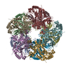 7v4hC 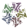 7v4jC  7v4kC 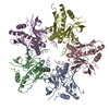 7v4lC M: map data used to model this data C: citing same article ( |
|---|---|
| Similar structure data |


-Links
 PDBj
PDBj
- Assembly
| Deposited unit | 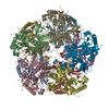
|
|---|---|
| 1 |
|
-Components
| #1: Protein | Mass: 39289.180 Da / Num. of mol.: 10 Source method: isolated from a genetically manipulated source Source: (gene. exp.) Camellia sinensis (black tea) / Gene: CsGS1;3, GS1.3 / Production host:  |
|---|
-Experimental details
-Experiment
| Experiment | Method: ELECTRON MICROSCOPY |
|---|---|
| EM experiment | Aggregation state: PARTICLE / 3D reconstruction method: single particle reconstruction |
- Sample preparation
| Component | Name: CsGS1b / Type: CELL / Entity ID: all / Source: RECOMBINANT |
|---|---|
| Source (natural) | Organism: |
| Source (recombinant) | Organism: |
| Buffer solution | pH: 7.4 |
| Specimen | Embedding applied: NO / Shadowing applied: NO / Staining applied: NO / Vitrification applied: YES |
| Vitrification | Cryogen name: ETHANE |
- Electron microscopy imaging
| Experimental equipment |  Model: Titan Krios / Image courtesy: FEI Company |
|---|---|
| Microscopy | Model: FEI TITAN KRIOS |
| Electron gun | Electron source:  FIELD EMISSION GUN / Accelerating voltage: 300 kV / Illumination mode: FLOOD BEAM FIELD EMISSION GUN / Accelerating voltage: 300 kV / Illumination mode: FLOOD BEAM |
| Electron lens | Mode: BRIGHT FIELD |
| Image recording | Electron dose: 51 e/Å2 / Film or detector model: GATAN K2 SUMMIT (4k x 4k) |
- Processing
| Software | Name: PHENIX / Version: 1.19_4092: / Classification: refinement | ||||||||||||||||||||||||
|---|---|---|---|---|---|---|---|---|---|---|---|---|---|---|---|---|---|---|---|---|---|---|---|---|---|
| CTF correction | Type: NONE | ||||||||||||||||||||||||
| 3D reconstruction | Resolution: 3.3 Å / Resolution method: FSC 0.143 CUT-OFF / Num. of particles: 43876 / Symmetry type: POINT | ||||||||||||||||||||||||
| Refine LS restraints |
|
