 Movie
Movie Controller

[English] 日本語
 Yorodumi
- PDB-7utd: The 2.19-angstrom CryoEM structure of the [NiFe]-hydrogenase Huc ... -
+ Open data

- Basic information
| Entry | Database: PDB / ID: 7utd | |||||||||
|---|---|---|---|---|---|---|---|---|---|---|
| Title | The 2.19-angstrom CryoEM structure of the [NiFe]-hydrogenase Huc from Mycobacterium smegmatis - Complex minus stalk | |||||||||
 Components Components |
| |||||||||
 Keywords Keywords | OXIDOREDUCTASE / [NiFe] Hydrogenase / Membrane associated / Complex / Quinone Transport / ELECTRON TRANSPORT / OXIDOREDUCTASE (EC 1.12.99.6) | |||||||||
| Function / homology |  Function and homology information Function and homology informationhydrogenase (acceptor) / ferredoxin hydrogenase complex / [Ni-Fe] hydrogenase complex / hydrogenase (acceptor) activity / ferredoxin hydrogenase activity / cell envelope / anaerobic respiration / 3 iron, 4 sulfur cluster binding / nickel cation binding / 4 iron, 4 sulfur cluster binding ...hydrogenase (acceptor) / ferredoxin hydrogenase complex / [Ni-Fe] hydrogenase complex / hydrogenase (acceptor) activity / ferredoxin hydrogenase activity / cell envelope / anaerobic respiration / 3 iron, 4 sulfur cluster binding / nickel cation binding / 4 iron, 4 sulfur cluster binding / electron transfer activity / membrane / metal ion binding Similarity search - Function | |||||||||
| Biological species |  Mycolicibacterium smegmatis MC2 155 (bacteria) Mycolicibacterium smegmatis MC2 155 (bacteria) | |||||||||
| Method | ELECTRON MICROSCOPY / single particle reconstruction / cryo EM / Resolution: 2.19 Å | |||||||||
 Authors Authors | Grinter, R. / Venugopal, H. / Kropp, A. / Greening, C. | |||||||||
| Funding support |  Australia, 2items Australia, 2items
| |||||||||
 Citation Citation | Journal: Nature / Year: 2023 Title: Structural basis for bacterial energy extraction from atmospheric hydrogen. Authors: Rhys Grinter / Ashleigh Kropp / Hari Venugopal / Moritz Senger / Jack Badley / Princess R Cabotaje / Ruyu Jia / Zehui Duan / Ping Huang / Sven T Stripp / Christopher K Barlow / Matthew ...Authors: Rhys Grinter / Ashleigh Kropp / Hari Venugopal / Moritz Senger / Jack Badley / Princess R Cabotaje / Ruyu Jia / Zehui Duan / Ping Huang / Sven T Stripp / Christopher K Barlow / Matthew Belousoff / Hannah S Shafaat / Gregory M Cook / Ralf B Schittenhelm / Kylie A Vincent / Syma Khalid / Gustav Berggren / Chris Greening /      Abstract: Diverse aerobic bacteria use atmospheric H as an energy source for growth and survival. This globally significant process regulates the composition of the atmosphere, enhances soil biodiversity and ...Diverse aerobic bacteria use atmospheric H as an energy source for growth and survival. This globally significant process regulates the composition of the atmosphere, enhances soil biodiversity and drives primary production in extreme environments. Atmospheric H oxidation is attributed to uncharacterized members of the [NiFe] hydrogenase superfamily. However, it remains unresolved how these enzymes overcome the extraordinary catalytic challenge of oxidizing picomolar levels of H amid ambient levels of the catalytic poison O and how the derived electrons are transferred to the respiratory chain. Here we determined the cryo-electron microscopy structure of the Mycobacterium smegmatis hydrogenase Huc and investigated its mechanism. Huc is a highly efficient oxygen-insensitive enzyme that couples oxidation of atmospheric H to the hydrogenation of the respiratory electron carrier menaquinone. Huc uses narrow hydrophobic gas channels to selectively bind atmospheric H at the expense of O, and 3 [3Fe-4S] clusters modulate the properties of the enzyme so that atmospheric H oxidation is energetically feasible. The Huc catalytic subunits form an octameric 833 kDa complex around a membrane-associated stalk, which transports and reduces menaquinone 94 Å from the membrane. These findings provide a mechanistic basis for the biogeochemically and ecologically important process of atmospheric H oxidation, uncover a mode of energy coupling dependent on long-range quinone transport, and pave the way for the development of catalysts that oxidize H in ambient air. | |||||||||
| History |
|
- Structure visualization
| Structure viewer | Molecule: MolmilJmol/JSmol |
|---|
- Downloads & links
-Download
| PDBx/mmCIF format | 7utd.cif.gz | 1.2 MB | Display | PDBx/mmCIF format |
|---|---|---|---|---|
| PDB format | pdb7utd.ent.gz | 970.7 KB | Display | PDB format |
| PDBx/mmJSON format | 7utd.json.gz | Tree view | PDBx/mmJSON format | |
| Others |  Other downloads Other downloads |
-Validation report
| Arichive directory | https://data.pdbj.org/pub/pdb/validation_reports/ut/7utdftp://data.pdbj.org/pub/pdb/validation_reports/ut/7utd | HTTPS FTP |
|---|
-Related structure data
| Related structure data | 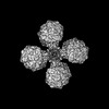 26767MC  7uurC  7uusC  8dqvC M: map data used to model this data C: citing same article ( |
|---|---|
| Similar structure data |

-Links
 PDBj
PDBj


- Assembly
| Deposited unit | 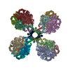
|
|---|---|
| 1 |
|
-Components
-Hydrogenase-2, ... , 2 types, 16 molecules ACEGIKMOBDFHJLNP
| #1: Protein | Mass: 57217.078 Da / Num. of mol.: 8 / Source method: isolated from a natural source Source: (natural) Mycolicibacterium smegmatis MC2 155 (bacteria)Strain: ATCC 700084 / mc(2)155 / References: UniProt: A0QUM7, hydrogenase (acceptor) #2: Protein | Mass: 39659.770 Da / Num. of mol.: 8 / Source method: isolated from a natural source Source: (natural) Mycolicibacterium smegmatis MC2 155 (bacteria)Strain: ATCC 700084 / mc(2)155 / References: UniProt: I7G634, hydrogenase (acceptor) |
|---|
-Protein , 1 types, 4 molecules QRST
| #3: Protein | Mass: 6416.264 Da / Num. of mol.: 4 / Source method: isolated from a natural source Source: (natural) Mycolicibacterium smegmatis MC2 155 (bacteria)Strain: MC2 155 / References: UniProt: A0QUM5 |
|---|
-Non-polymers , 5 types, 56 molecules 


| #4: Chemical | ChemComp-3NI /  Mass: 58.693 Da / Num. of mol.: 8 / Source method: obtained synthetically / Formula: Ni / Feature type: SUBJECT OF INVESTIGATION Mass: 58.693 Da / Num. of mol.: 8 / Source method: obtained synthetically / Formula: Ni / Feature type: SUBJECT OF INVESTIGATION#5: Chemical | ChemComp-FCO /  Mass: 135.890 Da / Num. of mol.: 8 / Source method: obtained synthetically / Formula: C3FeN2O / Feature type: SUBJECT OF INVESTIGATION Mass: 135.890 Da / Num. of mol.: 8 / Source method: obtained synthetically / Formula: C3FeN2O / Feature type: SUBJECT OF INVESTIGATION#6: Chemical | ChemComp-MG /  Mass: 24.305 Da / Num. of mol.: 8 / Source method: obtained synthetically / Formula: Mg Mass: 24.305 Da / Num. of mol.: 8 / Source method: obtained synthetically / Formula: Mg#7: Chemical | ChemComp-MQ9 /  Mass: 785.233 Da / Num. of mol.: 8 / Source method: obtained synthetically / Formula: C56H80O2 / Feature type: SUBJECT OF INVESTIGATION Mass: 785.233 Da / Num. of mol.: 8 / Source method: obtained synthetically / Formula: C56H80O2 / Feature type: SUBJECT OF INVESTIGATION#8: Chemical | ChemComp-F3S /  Mass: 295.795 Da / Num. of mol.: 24 / Source method: obtained synthetically / Formula: Fe3S4 / Feature type: SUBJECT OF INVESTIGATION Mass: 295.795 Da / Num. of mol.: 24 / Source method: obtained synthetically / Formula: Fe3S4 / Feature type: SUBJECT OF INVESTIGATION |
|---|
-Details
| Has ligand of interest | Y |
|---|---|
| Has protein modification | N |
-Experimental details
-Experiment
| Experiment | Method: ELECTRON MICROSCOPY |
|---|---|
| EM experiment | Aggregation state: PARTICLE / 3D reconstruction method: single particle reconstruction |
- Sample preparation
| Component | Name: Complex of the type 2 [NiFe]-hydrogenase Huc from Mycobacterium smegmatis Type: COMPLEX / Entity ID: #1-#3 / Source: NATURAL | |||||||||||||||
|---|---|---|---|---|---|---|---|---|---|---|---|---|---|---|---|---|
| Molecular weight | Value: 0.833 MDa / Experimental value: NO | |||||||||||||||
| Source (natural) | Organism: Mycolicibacterium smegmatis (bacteria) / Strain: MC2 155 | |||||||||||||||
| Buffer solution | pH: 7.9 / Details: pH 7.9 | |||||||||||||||
| Buffer component |
| |||||||||||||||
| Specimen | Conc.: 5 mg/ml / Embedding applied: NO / Shadowing applied: NO / Staining applied: NO / Vitrification applied: YES | |||||||||||||||
| Specimen support | Grid material: GOLD / Grid type: Quantifoil | |||||||||||||||
| Vitrification | Instrument: FEI VITROBOT MARK III / Cryogen name: ETHANE / Humidity: 100 % / Chamber temperature: 295 K |
- Electron microscopy imaging
| Experimental equipment |  Model: Titan Krios / Image courtesy: FEI Company |
|---|---|
| Microscopy | Model: FEI TITAN KRIOS |
| Electron gun | Electron source:  FIELD EMISSION GUN / Accelerating voltage: 300 kV / Illumination mode: FLOOD BEAM FIELD EMISSION GUN / Accelerating voltage: 300 kV / Illumination mode: FLOOD BEAM |
| Electron lens | Mode: BRIGHT FIELD / Nominal defocus max: 1500 nm / Nominal defocus min: 500 nm / Cs: 2.7 mm |
| Specimen holder | Cryogen: HELIUM |
| Image recording | Average exposure time: 6 sec. / Electron dose: 66 e/Å2 / Film or detector model: GATAN K3 (6k x 4k) / Num. of real images: 3113 |
- Processing
| EM software |
| ||||||||||||||||||||||||||||||||||||||||
|---|---|---|---|---|---|---|---|---|---|---|---|---|---|---|---|---|---|---|---|---|---|---|---|---|---|---|---|---|---|---|---|---|---|---|---|---|---|---|---|---|---|
| CTF correction | Type: PHASE FLIPPING AND AMPLITUDE CORRECTION | ||||||||||||||||||||||||||||||||||||||||
| Particle selection | Num. of particles selected: 825900 | ||||||||||||||||||||||||||||||||||||||||
| Symmetry | Point symmetry: C4 (4 fold cyclic) | ||||||||||||||||||||||||||||||||||||||||
| 3D reconstruction | Resolution: 2.19 Å / Resolution method: FSC 0.143 CUT-OFF / Num. of particles: 139367 / Num. of class averages: 1 / Symmetry type: POINT | ||||||||||||||||||||||||||||||||||||||||
| Atomic model building | Protocol: BACKBONE TRACE / Space: REAL / Target criteria: Correlation coefficient |
