 Movie
Movie Controller

[English] 日本語
 Yorodumi
- PDB-7usb: CCoV-HuPn-2018 S in the swung out conformation (local refinement ... -
+ Open data

- Basic information
| Entry | Database: PDB / ID: 7usb | ||||||
|---|---|---|---|---|---|---|---|
| Title | CCoV-HuPn-2018 S in the swung out conformation (local refinement of domain 0) | ||||||
 Components Components | Spike glycoprotein | ||||||
 Keywords Keywords | VIRAL PROTEIN / Human coronavirus / Coronavirus / CCoV-HuPn-2018 / spike glycoprotein / Alpha-coronaviruses / Structural Genomics / Seattle Structural Genomics Center for Infectious Disease / SSGCID | ||||||
| Function / homology |  Function and homology information Function and homology informationendocytosis involved in viral entry into host cell / host cell endoplasmic reticulum-Golgi intermediate compartment membrane / receptor-mediated virion attachment to host cell / fusion of virus membrane with host plasma membrane / fusion of virus membrane with host endosome membrane / viral envelope / virion membrane / membrane Similarity search - Function | ||||||
| Biological species |  unidentified human coronavirus unidentified human coronavirus | ||||||
| Method | ELECTRON MICROSCOPY / single particle reconstruction / cryo EM / Resolution: 3.1 Å | ||||||
 Authors Authors | Tortorici, M.A. / Veesler, D. / Seattle Structural Genomics Center for Infectious Disease (SSGCID) | ||||||
| Funding support |  United States, 1items United States, 1items
| ||||||
 Citation Citation | Journal: Cell / Year: 2022 Title: Structure, receptor recognition, and antigenicity of the human coronavirus CCoV-HuPn-2018 spike glycoprotein. Authors: M Alejandra Tortorici / Alexandra C Walls / Anshu Joshi / Young-Jun Park / Rachel T Eguia / Marcos C Miranda / Elizabeth Kepl / Annie Dosey / Terry Stevens-Ayers / Michael J Boeckh / Amalio ...Authors: M Alejandra Tortorici / Alexandra C Walls / Anshu Joshi / Young-Jun Park / Rachel T Eguia / Marcos C Miranda / Elizabeth Kepl / Annie Dosey / Terry Stevens-Ayers / Michael J Boeckh / Amalio Telenti / Antonio Lanzavecchia / Neil P King / Davide Corti / Jesse D Bloom / David Veesler /  Abstract: The isolation of CCoV-HuPn-2018 from a child respiratory swab indicates that more coronaviruses are spilling over to humans than previously appreciated. We determined the structures of the CCoV-HuPn- ...The isolation of CCoV-HuPn-2018 from a child respiratory swab indicates that more coronaviruses are spilling over to humans than previously appreciated. We determined the structures of the CCoV-HuPn-2018 spike glycoprotein trimer in two distinct conformational states and showed that its domain 0 recognizes sialosides. We identified that the CCoV-HuPn-2018 spike binds canine, feline, and porcine aminopeptidase N (APN) orthologs, which serve as entry receptors, and determined the structure of the receptor-binding B domain in complex with canine APN. The introduction of an oligosaccharide at position N739 of human APN renders cells susceptible to CCoV-HuPn-2018 spike-mediated entry, suggesting that single-nucleotide polymorphisms might account for viral detection in some individuals. Human polyclonal plasma antibodies elicited by HCoV-229E infection and a porcine coronavirus monoclonal antibody inhibit CCoV-HuPn-2018 spike-mediated entry, underscoring the cross-neutralizing activity among ɑ-coronaviruses. These data pave the way for vaccine and therapeutic development targeting this zoonotic pathogen representing the eighth human-infecting coronavirus. | ||||||
| History |
|
- Structure visualization
| Structure viewer | Molecule: MolmilJmol/JSmol |
|---|
- Downloads & links
-Download
| PDBx/mmCIF format | 7usb.cif.gz | 54.5 KB | Display | PDBx/mmCIF format |
|---|---|---|---|---|
| PDB format | pdb7usb.ent.gz | 38.1 KB | Display | PDB format |
| PDBx/mmJSON format | 7usb.json.gz | Tree view | PDBx/mmJSON format | |
| Others |  Other downloads Other downloads |
-Validation report
| Summary document | 7usb_validation.pdf.gz | 1.1 MB | Display | wwPDB validaton report |
|---|---|---|---|---|
| Full document | 7usb_full_validation.pdf.gz | 1.1 MB | Display | |
| Data in XML | 7usb_validation.xml.gz | 26.4 KB | Display | |
| Data in CIF | 7usb_validation.cif.gz | 34.1 KB | Display | |
| Arichive directory | https://data.pdbj.org/pub/pdb/validation_reports/us/7usbftp://data.pdbj.org/pub/pdb/validation_reports/us/7usb | HTTPS FTP |
-Related structure data
| Related structure data |  26731MC  7u0lC 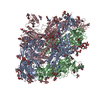 7us6C 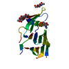 7us9C 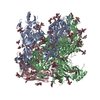 7usaC M: map data used to model this data C: citing same article ( |
|---|---|
| Similar structure data |


-Links
 PDBj
PDBj- Assembly
| Deposited unit | 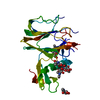
|
|---|---|
| 1 |
|
-Components
| #1: Protein | Mass: 37624.590 Da / Num. of mol.: 1 Fragment: CCoV-HuPn-2018 S domain 0 in swung out conformation Source method: isolated from a genetically manipulated source Source: (gene. exp.) unidentified human coronavirusProduction host:  References: UniProt: A0A8E6CMP0 | ||
|---|---|---|---|
| #2: Polysaccharide | 2-acetamido-2-deoxy-beta-D-glucopyranose-(1-4)-2-acetamido-2-deoxy-beta-D-glucopyranose Source method: isolated from a genetically manipulated source | ||
| #3: Sugar |   Type: D-saccharide, beta linking / Mass: 221.208 Da / Num. of mol.: 3 / Source method: obtained synthetically / Formula: C8H15NO6 Type: D-saccharide, beta linking / Mass: 221.208 Da / Num. of mol.: 3 / Source method: obtained synthetically / Formula: C8H15NO6Has ligand of interest | N | |
-Experimental details
-Experiment
| Experiment | Method: ELECTRON MICROSCOPY |
|---|---|
| EM experiment | Aggregation state: PARTICLE / 3D reconstruction method: single particle reconstruction |
- Sample preparation
| Component | Name: CCoV-HuPn-2018 / Type: VIRUS / Entity ID: #1 / Source: RECOMBINANT |
|---|---|
| Source (natural) | Organism:  CCoV-HuPn-2018 (virus) CCoV-HuPn-2018 (virus) |
| Source (recombinant) | Organism: |
| Details of virus | Empty: YES / Enveloped: YES / Isolate: OTHER / Type: VIRION |
| Natural host | Organism: Homo sapiens |
| Buffer solution | pH: 8 |
| Specimen | Embedding applied: YES / Shadowing applied: NO / Staining applied: NO / Vitrification applied: YES |
| EM embedding | Material: Tris buffer saline |
| Vitrification | Cryogen name: ETHANE |
- Electron microscopy imaging
| Experimental equipment |  Model: Titan Krios / Image courtesy: FEI Company |
|---|---|
| Microscopy | Model: FEI TITAN KRIOS |
| Electron gun | Electron source:  FIELD EMISSION GUN / Accelerating voltage: 300 kV / Illumination mode: FLOOD BEAM FIELD EMISSION GUN / Accelerating voltage: 300 kV / Illumination mode: FLOOD BEAM |
| Electron lens | Mode: BRIGHT FIELD / Nominal defocus max: 1500 nm / Nominal defocus min: 800 nm |
| Image recording | Electron dose: 80 e/Å2 / Film or detector model: GATAN K3 (6k x 4k) |
- Processing
| CTF correction | Type: PHASE FLIPPING AND AMPLITUDE CORRECTION |
|---|---|
| 3D reconstruction | Resolution: 3.1 Å / Resolution method: FSC 0.143 CUT-OFF / Num. of particles: 102954 / Symmetry type: POINT |
