 Movie
Movie Controller

[English] 日本語
 Yorodumi
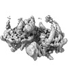
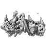
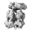
- PDB-7us2: PARL-cleaved Skd3 (human ClpB) E455Q Nucleotide Binding Domain he... -
+ Open data

- Basic information
| Entry | Database: PDB / ID: 7us2 | |||||||||||||||||||||
|---|---|---|---|---|---|---|---|---|---|---|---|---|---|---|---|---|---|---|---|---|---|---|
| Title | PARL-cleaved Skd3 (human ClpB) E455Q Nucleotide Binding Domain hexamer bound to ATPgammaS, open conformation | |||||||||||||||||||||
 Components Components |
| |||||||||||||||||||||
 Keywords Keywords | CHAPERONE / AAA+ ATPase / Mitochondria / Protein Folding | |||||||||||||||||||||
| Function / homology |  Function and homology information Function and homology informationgranulocyte differentiation / RIG-I signaling pathway / ATP-dependent protein disaggregase activity / Hydrolases; Acting on acid anhydrides; In phosphorus-containing anhydrides / antiviral innate immune response / mitochondrial intermembrane space / cellular response to heat / ATP hydrolysis activity / mitochondrion / ATP binding / cytoplasm Similarity search - Function | |||||||||||||||||||||
| Biological species |  Homo sapiens (human) Homo sapiens (human) | |||||||||||||||||||||
| Method | ELECTRON MICROSCOPY / single particle reconstruction / cryo EM / Resolution: 2.76 Å | |||||||||||||||||||||
 Authors Authors | Gupta, A. / Lentzsch, A.M. / Siegel, A.S. / Yu, Z. / Lu, C. / Chio, U.S. / Cheng, Y. / Shan, S.-o. | |||||||||||||||||||||
| Funding support |  United States, 6items United States, 6items
| |||||||||||||||||||||
 Citation Citation | Journal: Sci Adv / Year: 2023 Title: Dodecamer assembly of a metazoan AAA chaperone couples substrate extraction to refolding. Authors: Arpit Gupta / Alfred M Lentzsch / Alex Siegel / Zanlin Yu / Un Seng Chio / Yifan Cheng / Shu-Ou Shan / Abstract: Ring-forming AAA chaperones solubilize protein aggregates and protect organisms from proteostatic stress. In metazoans, the AAA chaperone Skd3 in the mitochondrial intermembrane space (IMS) is ...Ring-forming AAA chaperones solubilize protein aggregates and protect organisms from proteostatic stress. In metazoans, the AAA chaperone Skd3 in the mitochondrial intermembrane space (IMS) is critical for human health and efficiently refolds aggregated proteins, but its underlying mechanism is poorly understood. Here, we show that Skd3 harbors both disaggregase and protein refolding activities enabled by distinct assembly states. High-resolution structures of Skd3 hexamers in distinct conformations capture ratchet-like motions that mediate substrate extraction. Unlike previously described disaggregases, Skd3 hexamers further assemble into dodecameric cages in which solubilized substrate proteins can attain near-native states. Skd3 mutants defective in dodecamer assembly retain disaggregase activity but are impaired in client refolding, linking the disaggregase and refolding activities to the hexameric and dodecameric states of Skd3, respectively. We suggest that Skd3 is a combined disaggregase and foldase, and this property is particularly suited to meet the complex proteostatic demands in the mitochondrial IMS. | |||||||||||||||||||||
| History |
|
- Structure visualization
| Structure viewer | Molecule: MolmilJmol/JSmol |
|---|
- Downloads & links
-Download
| PDBx/mmCIF format | 7us2.cif.gz | 437.6 KB | Display | PDBx/mmCIF format |
|---|---|---|---|---|
| PDB format | pdb7us2.ent.gz | 350 KB | Display | PDB format |
| PDBx/mmJSON format | 7us2.json.gz | Tree view | PDBx/mmJSON format | |
| Others |  Other downloads Other downloads |
-Validation report
| Summary document | 7us2_validation.pdf.gz | 1.4 MB | Display | wwPDB validaton report |
|---|---|---|---|---|
| Full document | 7us2_full_validation.pdf.gz | 1.4 MB | Display | |
| Data in XML | 7us2_validation.xml.gz | 71.1 KB | Display | |
| Data in CIF | 7us2_validation.cif.gz | 102.8 KB | Display | |
| Arichive directory | https://data.pdbj.org/pub/pdb/validation_reports/us/7us2ftp://data.pdbj.org/pub/pdb/validation_reports/us/7us2 | HTTPS FTP |
-Related structure data
| Related structure data |  26722MC M: map data used to model this data C: citing same article ( |
|---|---|
| Similar structure data |
-Links
 PDBj
PDBj


- Assembly
| Deposited unit | 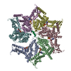
|
|---|---|
| 1 |
|
-Components
| #1: Protein | Mass: 65912.883 Da / Num. of mol.: 6 Source method: isolated from a genetically manipulated source Source: (gene. exp.) Homo sapiens (human) / Gene: CLPB, HSP78, SKD3 / Production host:  References: UniProt: Q9H078, Hydrolases; Acting on acid anhydrides; In phosphorus-containing anhydrides #2: Protein/peptide | | Mass: 1209.482 Da / Num. of mol.: 1 Source method: isolated from a genetically manipulated source Source: (gene. exp.) Homo sapiens (human) / Production host: #3: Chemical | ChemComp-AGS /   Mass: 523.247 Da / Num. of mol.: 6 / Source method: obtained synthetically / Formula: C10H16N5O12P3S / Feature type: SUBJECT OF INVESTIGATION / Comment: ATP-gamma-S, energy-carrying molecule analogue*YM Mass: 523.247 Da / Num. of mol.: 6 / Source method: obtained synthetically / Formula: C10H16N5O12P3S / Feature type: SUBJECT OF INVESTIGATION / Comment: ATP-gamma-S, energy-carrying molecule analogue*YM#4: Chemical | ChemComp-MG /   Mass: 24.305 Da / Num. of mol.: 6 / Source method: obtained synthetically / Formula: Mg / Feature type: SUBJECT OF INVESTIGATION Mass: 24.305 Da / Num. of mol.: 6 / Source method: obtained synthetically / Formula: Mg / Feature type: SUBJECT OF INVESTIGATIONHas ligand of interest | Y | |
|---|
-Experimental details
-Experiment
| Experiment | Method: ELECTRON MICROSCOPY |
|---|---|
| EM experiment | Aggregation state: PARTICLE / 3D reconstruction method: single particle reconstruction |
- Sample preparation
| Component | Name: PARL-cleaved Skd3 / Type: COMPLEX / Entity ID: #1-#2 / Source: RECOMBINANT |
|---|---|
| Molecular weight | Value: 0.397 MDa / Experimental value: NO |
| Source (natural) | Organism: Homo sapiens (human) |
| Source (recombinant) | Organism: |
| Buffer solution | pH: 7.4 |
| Specimen | Conc.: 0.1 mg/ml / Embedding applied: NO / Shadowing applied: NO / Staining applied: NO / Vitrification applied: YES |
| Vitrification | Cryogen name: ETHANE / Humidity: 100 % |
- Electron microscopy imaging
| Experimental equipment |  Model: Titan Krios / Image courtesy: FEI Company |
|---|---|
| Microscopy | Model: FEI TITAN KRIOS |
| Electron gun | Electron source:  FIELD EMISSION GUN / Accelerating voltage: 300 kV / Illumination mode: FLOOD BEAM FIELD EMISSION GUN / Accelerating voltage: 300 kV / Illumination mode: FLOOD BEAM |
| Electron lens | Mode: BRIGHT FIELD / Nominal defocus max: -2000 nm / Nominal defocus min: -1000 nm |
| Image recording | Electron dose: 60 e/Å2 / Film or detector model: FEI FALCON IV (4k x 4k) |
- Processing
| CTF correction | Type: NONE |
|---|---|
| 3D reconstruction | Resolution: 2.76 Å / Resolution method: FSC 0.143 CUT-OFF / Num. of particles: 1459443 / Symmetry type: POINT |
