 Movie
Movie Controller

[English] 日本語
 Yorodumi
- PDB-7um0: Structure of the phage AR9 non-virion RNA polymerase holoenzyme i... -
+ Open data

- Basic information
| Entry | Database: PDB / ID: 7um0 | |||||||||
|---|---|---|---|---|---|---|---|---|---|---|
| Title | Structure of the phage AR9 non-virion RNA polymerase holoenzyme in complex with two DNA oligonucleotides containing the AR9 P077 promoter as determined by cryo-EM | |||||||||
 Components Components |
| |||||||||
 Keywords Keywords | TRANSCRIPTION / RNAP / deoxyuridine / template-strand promoter / sigma-like factor / gp226 | |||||||||
| Function / homology |  Function and homology information Function and homology informationDNA-directed RNA polymerase complex / ribonucleoside binding / DNA-directed RNA polymerase / DNA-directed RNA polymerase activity / DNA-templated transcription / DNA binding / metal ion binding Similarity search - Function | |||||||||
| Biological species |  Bacillus phage AR9 (virus) Bacillus phage AR9 (virus)synthetic construct (others) | |||||||||
| Method | ELECTRON MICROSCOPY / single particle reconstruction / cryo EM / Resolution: 3.8 Å | |||||||||
 Authors Authors | Leiman, P.G. / Fraser, A. / Sokolova, M.L. | |||||||||
| Funding support |  United States, 1items United States, 1items
| |||||||||
 Citation Citation | Journal: Nat Commun / Year: 2022 Title: Structural basis of template strand deoxyuridine promoter recognition by a viral RNA polymerase. Authors: Alec Fraser / Maria L Sokolova / Arina V Drobysheva / Julia V Gordeeva / Sergei Borukhov / John Jumper / Konstantin V Severinov / Petr G Leiman /   Abstract: Recognition of promoters in bacterial RNA polymerases (RNAPs) is controlled by sigma subunits. The key sequence motif recognized by the sigma, the -10 promoter element, is located in the non-template ...Recognition of promoters in bacterial RNA polymerases (RNAPs) is controlled by sigma subunits. The key sequence motif recognized by the sigma, the -10 promoter element, is located in the non-template strand of the double-stranded DNA molecule ~10 nucleotides upstream of the transcription start site. Here, we explain the mechanism by which the phage AR9 non-virion RNAP (nvRNAP), a bacterial RNAP homolog, recognizes the -10 element of its deoxyuridine-containing promoter in the template strand. The AR9 sigma-like subunit, the nvRNAP enzyme core, and the template strand together form two nucleotide base-accepting pockets whose shapes dictate the requirement for the conserved deoxyuridines. A single amino acid substitution in the AR9 sigma-like subunit allows one of these pockets to accept a thymine thus expanding the promoter consensus. Our work demonstrates the extent to which viruses can evolve host-derived multisubunit enzymes to make transcription of their own genes independent of the host. | |||||||||
| History |
|
- Structure visualization
| Structure viewer | Molecule: MolmilJmol/JSmol |
|---|
- Downloads & links
-Download
| PDBx/mmCIF format | 7um0.cif.gz | 584.3 KB | Display | PDBx/mmCIF format |
|---|---|---|---|---|
| PDB format | pdb7um0.ent.gz | 392.6 KB | Display | PDB format |
| PDBx/mmJSON format | 7um0.json.gz | Tree view | PDBx/mmJSON format | |
| Others |  Other downloads Other downloads |
-Validation report
| Arichive directory | https://data.pdbj.org/pub/pdb/validation_reports/um/7um0ftp://data.pdbj.org/pub/pdb/validation_reports/um/7um0 | HTTPS FTP |
|---|
-Related structure data
| Related structure data |  24763MC 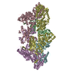 7s00C 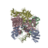 7s01C 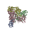 7um1C M: map data used to model this data C: citing same article ( |
|---|---|
| Similar structure data |
-Links
 PDBj
PDBj
- Assembly
| Deposited unit | 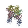
|
|---|---|
| 1 |
|
-Components
-DNA-directed RNA ... , 5 types, 5 molecules AdcDC
| #1: Protein | Mass: 54917.449 Da / Num. of mol.: 1 Source method: isolated from a genetically manipulated source Details: Promoter-specificity subunit / Source: (gene. exp.) Bacillus phage AR9 (virus) / Gene: AR9_g226 / Production host:  |
|---|---|
| #2: Protein | Mass: 51947.844 Da / Num. of mol.: 1 Source method: isolated from a genetically manipulated source Details: N-terminal part of beta-prime subunit / Source: (gene. exp.) Bacillus phage AR9 (virus) / Gene: AR9_g270 / Production host: |
| #3: Protein | Mass: 58112.289 Da / Num. of mol.: 1 Source method: isolated from a genetically manipulated source Details: N-terminal part of beta subunit / Source: (gene. exp.) Bacillus phage AR9 (virus) / Gene: AR9_g105 / Variant: Star / Production host: |
| #4: Protein | Mass: 73100.977 Da / Num. of mol.: 1 Source method: isolated from a genetically manipulated source Details: C-terminal part of beta-prime subunit / Source: (gene. exp.) Bacillus phage AR9 (virus) / Gene: AR9_g154 / Production host: References: UniProt: A0A172JI62, DNA-directed RNA polymerase |
| #5: Protein | Mass: 76978.383 Da / Num. of mol.: 1 Source method: isolated from a genetically manipulated source Details: C-terminal part of beta subunit / Source: (gene. exp.) Bacillus phage AR9 (virus) / Gene: AR9_g089 / Production host: References: UniProt: A0A172JHZ2, DNA-directed RNA polymerase |
-DNA chain / Non-polymers , 2 types, 2 molecules B
| #6: DNA chain | Mass: 864.582 Da / Num. of mol.: 1 / Source method: obtained synthetically / Source: (synth.) synthetic construct (others) |
|---|---|
| #7: Chemical | ChemComp-ZN /  Mass: 65.409 Da / Num. of mol.: 1 / Source method: obtained synthetically / Formula: Zn Mass: 65.409 Da / Num. of mol.: 1 / Source method: obtained synthetically / Formula: Zn |
-Details
| Has ligand of interest | Y |
|---|
-Experimental details
-Experiment
| Experiment | Method: ELECTRON MICROSCOPY |
|---|---|
| EM experiment | Aggregation state: PARTICLE / 3D reconstruction method: single particle reconstruction |
- Sample preparation
| Component | Name: AR9 nvRNAP promoter complex / Type: COMPLEX Details: The complex consists of the AR9 nvRNAP holoenzyme and two copies of the same oligonucleotide containing the P077 promoter. Only three bases in one of the oligonucleotides are sufficiently ...Details: The complex consists of the AR9 nvRNAP holoenzyme and two copies of the same oligonucleotide containing the P077 promoter. Only three bases in one of the oligonucleotides are sufficiently ordered for atomic model building. Entity ID: #1-#5 / Source: RECOMBINANT |
|---|---|
| Molecular weight | Experimental value: NO |
| Source (natural) | Organism: Bacillus phage AR9 (virus) |
| Source (recombinant) | Organism: |
| Buffer solution | pH: 6.8 |
| Specimen | Conc.: 10 mg/ml / Embedding applied: NO / Shadowing applied: NO / Staining applied: NO / Vitrification applied: YES |
| Specimen support | Grid material: COPPER / Grid type: Quantifoil R1.2/1.3 |
| Vitrification | Cryogen name: ETHANE |
- Electron microscopy imaging
| Experimental equipment |  Model: Titan Krios / Image courtesy: FEI Company |
|---|---|
| Microscopy | Model: FEI TITAN KRIOS |
| Electron gun | Electron source:  FIELD EMISSION GUN / Accelerating voltage: 300 kV / Illumination mode: FLOOD BEAM FIELD EMISSION GUN / Accelerating voltage: 300 kV / Illumination mode: FLOOD BEAM |
| Electron lens | Mode: BRIGHT FIELD / Nominal defocus max: 4000 nm / Nominal defocus min: 1000 nm |
| Image recording | Electron dose: 44 e/Å2 / Film or detector model: GATAN K3 BIOQUANTUM (6k x 4k) |
- Processing
| Software |
| ||||||||||||||||||||||||
|---|---|---|---|---|---|---|---|---|---|---|---|---|---|---|---|---|---|---|---|---|---|---|---|---|---|
| EM software |
| ||||||||||||||||||||||||
| CTF correction | Type: PHASE FLIPPING AND AMPLITUDE CORRECTION | ||||||||||||||||||||||||
| 3D reconstruction | Resolution: 3.8 Å / Resolution method: FSC 0.143 CUT-OFF / Num. of particles: 106867 / Symmetry type: POINT | ||||||||||||||||||||||||
| Atomic model building | Protocol: AB INITIO MODEL / Space: REAL | ||||||||||||||||||||||||
| Atomic model building | PDB-ID: 7S01 Accession code: 7S01 / Source name: PDB / Type: experimental model | ||||||||||||||||||||||||
| Refinement | Cross valid method: NONE Stereochemistry target values: GeoStd + Monomer Library + CDL v1.2 | ||||||||||||||||||||||||
| Displacement parameters | Biso mean: 88.16 Å2 | ||||||||||||||||||||||||
| Refine LS restraints |
|
