[English] 日本語
 Yorodumi
- PDB-7um1: Structure of bacteriophage AR9 non-virion RNAP polymerase holoenz... -
+ Open data
- Basic information
| Entry | Database: PDB / ID: 7um1 | ||||||
|---|---|---|---|---|---|---|---|
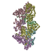
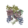
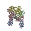
| Title | Structure of bacteriophage AR9 non-virion RNAP polymerase holoenzyme determined by cryo-EM | ||||||
 Components Components | (DNA-directed RNA ...) x 5 | ||||||
 Keywords Keywords | TRANSCRIPTION / RNAP / deoxyuridine / template-strand promoter / sigma-like factor / gp226 | ||||||
| Function / homology |  Function and homology information Function and homology informationDNA-directed RNA polymerase complex / ribonucleoside binding / DNA-directed RNA polymerase / DNA-directed RNA polymerase activity / DNA-templated transcription / DNA binding / metal ion binding Similarity search - Function | ||||||
| Biological species |  Bacillus phage AR9 (virus) Bacillus phage AR9 (virus) | ||||||
| Method | ELECTRON MICROSCOPY / single particle reconstruction / cryo EM / Resolution: 4.2 Å | ||||||
 Authors Authors | Leiman, P.G. / Fraser, A. / Sokolova, M.L. | ||||||
| Funding support |  United States, 1items United States, 1items
| ||||||
 Citation Citation |  Journal: Nat Commun / Year: 2022 Journal: Nat Commun / Year: 2022Title: Structural basis of template strand deoxyuridine promoter recognition by a viral RNA polymerase. Authors: Alec Fraser / Maria L Sokolova / Arina V Drobysheva / Julia V Gordeeva / Sergei Borukhov / John Jumper / Konstantin V Severinov / Petr G Leiman /    Abstract: Recognition of promoters in bacterial RNA polymerases (RNAPs) is controlled by sigma subunits. The key sequence motif recognized by the sigma, the -10 promoter element, is located in the non-template ...Recognition of promoters in bacterial RNA polymerases (RNAPs) is controlled by sigma subunits. The key sequence motif recognized by the sigma, the -10 promoter element, is located in the non-template strand of the double-stranded DNA molecule ~10 nucleotides upstream of the transcription start site. Here, we explain the mechanism by which the phage AR9 non-virion RNAP (nvRNAP), a bacterial RNAP homolog, recognizes the -10 element of its deoxyuridine-containing promoter in the template strand. The AR9 sigma-like subunit, the nvRNAP enzyme core, and the template strand together form two nucleotide base-accepting pockets whose shapes dictate the requirement for the conserved deoxyuridines. A single amino acid substitution in the AR9 sigma-like subunit allows one of these pockets to accept a thymine thus expanding the promoter consensus. Our work demonstrates the extent to which viruses can evolve host-derived multisubunit enzymes to make transcription of their own genes independent of the host. | ||||||
| History |
|
- Structure visualization
| Structure viewer | Molecule:  Molmil Molmil Jmol/JSmol Jmol/JSmol |
|---|
- Downloads & links
- Download
| PDBx/mmCIF format |  7um1.cif.gz 7um1.cif.gz | 501.8 KB | Display |  PDBx/mmCIF format PDBx/mmCIF format |
|---|---|---|---|---|
| PDB format |  pdb7um1.ent.gz pdb7um1.ent.gz | 334.4 KB | Display |  PDB format PDB format |
| PDBx/mmJSON format |  7um1.json.gz 7um1.json.gz | Tree view |  PDBx/mmJSON format PDBx/mmJSON format | |
| Others |  Other downloads Other downloads |
-Validation report
| Arichive directory |  https://data.pdbj.org/pub/pdb/validation_reports/um/7um1 https://data.pdbj.org/pub/pdb/validation_reports/um/7um1 ftp://data.pdbj.org/pub/pdb/validation_reports/um/7um1 ftp://data.pdbj.org/pub/pdb/validation_reports/um/7um1 | HTTPS FTP |
|---|
-Related structure data
| Related structure data |  24765MC  7s00C  7s01C  7um0C M: map data used to model this data C: citing same article ( |
|---|---|
| Similar structure data | Similarity search - Function & homology  F&H Search F&H Search |
- Links
Links
- Assembly
| Deposited unit | 
|
|---|---|
| 1 |
|
- Components
-DNA-directed RNA ... , 5 types, 5 molecules AdcDC
| #1: Protein | Mass: 54917.449 Da / Num. of mol.: 1 Source method: isolated from a genetically manipulated source Details: Promoter-specificity subunit / Source: (gene. exp.)  Bacillus phage AR9 (virus) / Gene: AR9_g226 / Production host: Bacillus phage AR9 (virus) / Gene: AR9_g226 / Production host:  |
|---|---|
| #2: Protein | Mass: 51947.844 Da / Num. of mol.: 1 / Mutation: N-terminal His-tag Source method: isolated from a genetically manipulated source Details: N-terminal part of beta-prime subunit / Source: (gene. exp.)  Bacillus phage AR9 (virus) / Gene: AR9_g270 / Production host: Bacillus phage AR9 (virus) / Gene: AR9_g270 / Production host:  |
| #3: Protein | Mass: 58112.289 Da / Num. of mol.: 1 Source method: isolated from a genetically manipulated source Details: N-terminal part of beta subunit / Source: (gene. exp.)  Bacillus phage AR9 (virus) / Gene: AR9_g105 / Production host: Bacillus phage AR9 (virus) / Gene: AR9_g105 / Production host:  |
| #4: Protein | Mass: 73100.977 Da / Num. of mol.: 1 Source method: isolated from a genetically manipulated source Details: C-terminal part of beta-prime subunit / Source: (gene. exp.)  Bacillus phage AR9 (virus) / Gene: AR9_g154 / Production host: Bacillus phage AR9 (virus) / Gene: AR9_g154 / Production host:  References: UniProt: A0A172JI62, DNA-directed RNA polymerase |
| #5: Protein | Mass: 76978.383 Da / Num. of mol.: 1 Source method: isolated from a genetically manipulated source Details: C-terminal part of beta subunit / Source: (gene. exp.)  Bacillus phage AR9 (virus) / Gene: AR9_g089 / Production host: Bacillus phage AR9 (virus) / Gene: AR9_g089 / Production host:  References: UniProt: A0A172JHZ2, DNA-directed RNA polymerase |
-Non-polymers , 1 types, 1 molecules 
| #6: Chemical | ChemComp-ZN / |
|---|
-Details
| Has ligand of interest | N |
|---|
-Experimental details
-Experiment
| Experiment | Method: ELECTRON MICROSCOPY |
|---|---|
| EM experiment | Aggregation state: PARTICLE / 3D reconstruction method: single particle reconstruction |
- Sample preparation
| Component | Name: Bacteriophage AR9 non-virion RNA polymerase holoenzyme Type: COMPLEX / Entity ID: #1-#5 / Source: RECOMBINANT |
|---|---|
| Molecular weight | Experimental value: NO |
| Source (natural) | Organism:  Bacillus phage AR9 (virus) Bacillus phage AR9 (virus) |
| Source (recombinant) | Organism:  |
| Buffer solution | pH: 6.8 |
| Specimen | Conc.: 20 mg/ml / Embedding applied: NO / Shadowing applied: NO / Staining applied: NO / Vitrification applied: YES |
| Vitrification | Cryogen name: ETHANE |
- Electron microscopy imaging
| Experimental equipment |  Model: Titan Krios / Image courtesy: FEI Company |
|---|---|
| Microscopy | Model: FEI TITAN KRIOS |
| Electron gun | Electron source:  FIELD EMISSION GUN / Accelerating voltage: 300 kV / Illumination mode: FLOOD BEAM FIELD EMISSION GUN / Accelerating voltage: 300 kV / Illumination mode: FLOOD BEAM |
| Electron lens | Mode: BRIGHT FIELD / Nominal defocus max: 2400 nm / Nominal defocus min: 1200 nm |
| Image recording | Electron dose: 43.2 e/Å2 / Film or detector model: GATAN K2 SUMMIT (4k x 4k) |
- Processing
| Software |
| ||||||||||||||||||||||||
|---|---|---|---|---|---|---|---|---|---|---|---|---|---|---|---|---|---|---|---|---|---|---|---|---|---|
| EM software |
| ||||||||||||||||||||||||
| CTF correction | Type: PHASE FLIPPING AND AMPLITUDE CORRECTION | ||||||||||||||||||||||||
| 3D reconstruction | Resolution: 4.2 Å / Resolution method: FSC 0.143 CUT-OFF / Num. of particles: 104471 / Symmetry type: POINT | ||||||||||||||||||||||||
| Atomic model building | Protocol: OTHER | ||||||||||||||||||||||||
| Atomic model building | PDB-ID: 7S01 Accession code: 7S01 / Source name: PDB / Type: experimental model | ||||||||||||||||||||||||
| Refinement | Cross valid method: NONE Stereochemistry target values: GeoStd + Monomer Library + CDL v1.2 | ||||||||||||||||||||||||
| Displacement parameters | Biso mean: 120.22 Å2 | ||||||||||||||||||||||||
| Refine LS restraints |
|
 Movie
Movie Controller


 PDBj
PDBj

