 Movie
Movie Controller


+ Open data

- Basic information
| Entry | Database: PDB / ID: 7u5s | ||||||
|---|---|---|---|---|---|---|---|
| Title | CryoEM structure of the Candida albicans Aro1 dimer | ||||||
 Components Components | Pentafunctional AROM polypeptide | ||||||
 Keywords Keywords | BIOSYNTHETIC PROTEIN | ||||||
| Function / homology |  Function and homology information Function and homology informationaromatic amino acid transport / 3-dehydroquinate synthase / 3-dehydroquinate synthase activity / shikimate kinase / shikimate kinase activity / shikimate dehydrogenase (NADP+) / shikimate 3-dehydrogenase (NADP+) activity / 3-phosphoshikimate 1-carboxyvinyltransferase / 3-phosphoshikimate 1-carboxyvinyltransferase activity / 3-dehydroquinate dehydratase ...aromatic amino acid transport / 3-dehydroquinate synthase / 3-dehydroquinate synthase activity / shikimate kinase / shikimate kinase activity / shikimate dehydrogenase (NADP+) / shikimate 3-dehydrogenase (NADP+) activity / 3-phosphoshikimate 1-carboxyvinyltransferase / 3-phosphoshikimate 1-carboxyvinyltransferase activity / 3-dehydroquinate dehydratase / 3-dehydroquinate dehydratase activity / chorismate biosynthetic process / aromatic amino acid biosynthetic process / amino acid biosynthetic process / cellular response to xenobiotic stimulus / ATP binding / metal ion binding / cytoplasm Similarity search - Function | ||||||
| Biological species |  Candida albicans (yeast) Candida albicans (yeast) | ||||||
| Method | ELECTRON MICROSCOPY / single particle reconstruction / cryo EM / Resolution: 4.16 Å | ||||||
 Authors Authors | Quade, B. / Borek, D. / Otwinowski, Z. / Center for Structural Genomics of Infectious Diseases (CSGID) | ||||||
| Funding support |  United States, 1items United States, 1items
| ||||||
 Citation Citation | Journal: Life Sci Alliance / Year: 2022 Title: Molecular analysis and essentiality of Aro1 shikimate biosynthesis multi-enzyme in . Authors: Peter J Stogios / Sean D Liston / Cameron Semper / Bradley Quade / Karolina Michalska / Elena Evdokimova / Shane Ram / Zbyszek Otwinowski / Dominika Borek / Leah E Cowen / Alexei Savchenko /  Abstract: In the human fungal pathogen , encodes an essential multi-enzyme that catalyses consecutive steps in the shikimate pathway for biosynthesis of chorismate, a precursor to folate and the aromatic ...In the human fungal pathogen , encodes an essential multi-enzyme that catalyses consecutive steps in the shikimate pathway for biosynthesis of chorismate, a precursor to folate and the aromatic amino acids. We obtained the first molecular image of Aro1 that reveals the architecture of all five enzymatic domains and their arrangement in the context of the full-length protein. Aro1 forms a flexible dimer allowing relative autonomy of enzymatic function of the individual domains. Our activity and in cellulo data suggest that only four of Aro1's enzymatic domains are functional and essential for viability of , whereas the 3-dehydroquinate dehydratase (DHQase) domain is inactive because of active site substitutions. We further demonstrate that in , the type II DHQase Dqd1 can compensate for the inactive DHQase domain of Aro1, suggesting an unrecognized essential role for this enzyme in shikimate biosynthesis. In contrast, in and , which do not encode a Dqd1 homolog, Aro1 DHQase domains are enzymatically active, highlighting diversity across species. | ||||||
| History |
|
- Structure visualization
| Structure viewer | Molecule: MolmilJmol/JSmol |
|---|
- Downloads & links
-Download
| PDBx/mmCIF format | 7u5s.cif.gz | 751.4 KB | Display | PDBx/mmCIF format |
|---|---|---|---|---|
| PDB format | pdb7u5s.ent.gz | 586.8 KB | Display | PDB format |
| PDBx/mmJSON format | 7u5s.json.gz | Tree view | PDBx/mmJSON format | |
| Others |  Other downloads Other downloads |
-Validation report
| Arichive directory | https://data.pdbj.org/pub/pdb/validation_reports/u5/7u5sftp://data.pdbj.org/pub/pdb/validation_reports/u5/7u5s | HTTPS FTP |
|---|
-Related structure data
| Related structure data |  26357MC  6c5cC  7tbuC 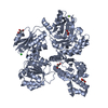 7tbvC  7u5tC  7u5uC M: map data used to model this data C: citing same article ( |
|---|---|
| Similar structure data |

-Links
 PDBj
PDBj

- Assembly
| Deposited unit | 
|
|---|---|
| 1 |
|
-Components
| #1: Protein | Mass: 169596.250 Da / Num. of mol.: 2 / Source method: isolated from a natural source / Source: (natural) Candida albicans (yeast) / Strain: SC5314 / ATCC MYA-2876References: UniProt: Q5AME2, 3-dehydroquinate synthase, 3-phosphoshikimate 1-carboxyvinyltransferase, shikimate kinase, 3-dehydroquinate dehydratase, shikimate dehydrogenase (NADP+) |
|---|
-Experimental details
-Experiment
| Experiment | Method: ELECTRON MICROSCOPY |
|---|---|
| EM experiment | Aggregation state: PARTICLE / 3D reconstruction method: single particle reconstruction |
- Sample preparation
| Component | Name: Aro1 dimer from Candida albicans / Type: COMPLEX / Entity ID: all / Source: NATURAL | |||||||||||||||
|---|---|---|---|---|---|---|---|---|---|---|---|---|---|---|---|---|
| Molecular weight | Value: 0.1694 MDa / Experimental value: NO | |||||||||||||||
| Source (natural) | Organism: Candida albicans (yeast) / Strain: CaLC6830 | |||||||||||||||
| Buffer solution | pH: 7.5 | |||||||||||||||
| Buffer component |
| |||||||||||||||
| Specimen | Conc.: 2.2 mg/ml / Embedding applied: NO / Shadowing applied: NO / Staining applied: NO / Vitrification applied: YES | |||||||||||||||
| Specimen support | Grid material: GOLD / Grid mesh size: 300 divisions/in. / Grid type: Quantifoil R1.2/1.3 | |||||||||||||||
| Vitrification | Instrument: FEI VITROBOT MARK IV / Cryogen name: ETHANE / Humidity: 100 % / Chamber temperature: 277 K |
- Electron microscopy imaging
| Experimental equipment |  Model: Titan Krios / Image courtesy: FEI Company |
|---|---|
| Microscopy | Model: FEI TITAN KRIOS |
| Electron gun | Electron source:  FIELD EMISSION GUN / Accelerating voltage: 300 kV / Illumination mode: FLOOD BEAM FIELD EMISSION GUN / Accelerating voltage: 300 kV / Illumination mode: FLOOD BEAM |
| Electron lens | Mode: BRIGHT FIELD / Nominal magnification: 105000 X / Nominal defocus max: 3000 nm / Nominal defocus min: 1500 nm / Cs: 2.7 mm / Alignment procedure: COMA FREE |
| Specimen holder | Cryogen: NITROGEN / Specimen holder model: FEI TITAN KRIOS AUTOGRID HOLDER |
| Image recording | Electron dose: 83.5 e/Å2 / Film or detector model: GATAN K3 (6k x 4k) |
- Processing
| EM software |
| ||||||||||||||||||||||||||||
|---|---|---|---|---|---|---|---|---|---|---|---|---|---|---|---|---|---|---|---|---|---|---|---|---|---|---|---|---|---|
| CTF correction | Type: PHASE FLIPPING AND AMPLITUDE CORRECTION | ||||||||||||||||||||||||||||
| Particle selection | Num. of particles selected: 1312555 | ||||||||||||||||||||||||||||
| Symmetry | Point symmetry: C1 (asymmetric) | ||||||||||||||||||||||||||||
| 3D reconstruction | Resolution: 4.16 Å / Resolution method: FSC 0.143 CUT-OFF / Num. of particles: 87484 / Symmetry type: POINT | ||||||||||||||||||||||||||||
| Atomic model building | Protocol: RIGID BODY FIT / Space: REAL | ||||||||||||||||||||||||||||
| Atomic model building |
|
