Movie
Movie Controller

[English] 日本語
 Yorodumi
- PDB-7tyr: Cryo-EM structure of the basal state of the Artemis:DNA-PKcs comp... -
+ Open data

- Basic information
| Entry | Database: PDB / ID: 7tyr | |||||||||
|---|---|---|---|---|---|---|---|---|---|---|
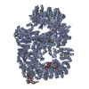
| Title | Cryo-EM structure of the basal state of the Artemis:DNA-PKcs complex (see COMPND 13/14) | |||||||||
 Components Components |
| |||||||||
 Keywords Keywords | DNA BINDING PROTEIN / Kinase / nuclease | |||||||||
| Function / homology |  Function and homology information Function and homology informationpositive regulation of platelet formation / single-stranded DNA endodeoxyribonuclease activity / T cell receptor V(D)J recombination / pro-B cell differentiation / DNA-dependent protein kinase activity / small-subunit processome assembly / positive regulation of lymphocyte differentiation / histone H2AXS139 kinase activity / DNA-dependent protein kinase complex / DNA-dependent protein kinase-DNA ligase 4 complex ...positive regulation of platelet formation / single-stranded DNA endodeoxyribonuclease activity / T cell receptor V(D)J recombination / pro-B cell differentiation / DNA-dependent protein kinase activity / small-subunit processome assembly / positive regulation of lymphocyte differentiation / histone H2AXS139 kinase activity / DNA-dependent protein kinase complex / DNA-dependent protein kinase-DNA ligase 4 complex / immunoglobulin V(D)J recombination / nonhomologous end joining complex / immature B cell differentiation / V(D)J recombination / regulation of smooth muscle cell proliferation / double-strand break repair via alternative nonhomologous end joining / regulation of epithelial cell proliferation / Cytosolic sensors of pathogen-associated DNA / telomere capping / IRF3-mediated induction of type I IFN / 5'-3' exonuclease activity / regulation of hematopoietic stem cell differentiation / 5'-3' DNA exonuclease activity / U3 snoRNA binding / T cell lineage commitment / maturation of 5.8S rRNA / positive regulation of double-strand break repair via nonhomologous end joining / negative regulation of cGAS/STING signaling pathway / B cell lineage commitment / response to ionizing radiation / peptidyl-threonine phosphorylation / negative regulation of protein phosphorylation / somitogenesis / ectopic germ cell programmed cell death / interstrand cross-link repair / mitotic G1 DNA damage checkpoint signaling / activation of innate immune response / telomere maintenance / positive regulation of erythrocyte differentiation / B cell differentiation / positive regulation of translation / response to gamma radiation / Nonhomologous End-Joining (NHEJ) / protein modification process / small-subunit processome / peptidyl-serine phosphorylation / protein-DNA complex / regulation of circadian rhythm / brain development / double-strand break repair via nonhomologous end joining / protein destabilization / cellular response to insulin stimulus / intrinsic apoptotic signaling pathway in response to DNA damage / T cell differentiation in thymus / rhythmic process / double-strand break repair / E3 ubiquitin ligases ubiquitinate target proteins / heart development / double-stranded DNA binding / endonuclease activity / transcription regulator complex / damaged DNA binding / Hydrolases; Acting on ester bonds / RNA polymerase II-specific DNA-binding transcription factor binding / adaptive immune response / protein phosphorylation / chromosome, telomeric region / protein kinase activity / non-specific serine/threonine protein kinase / positive regulation of apoptotic process / protein domain specific binding / innate immune response / protein serine kinase activity / protein serine/threonine kinase activity / DNA damage response / negative regulation of apoptotic process / chromatin / nucleolus / enzyme binding / Golgi apparatus / positive regulation of transcription by RNA polymerase II / protein-containing complex / RNA binding / nucleoplasm / ATP binding / membrane / nucleus / cytosol Similarity search - Function | |||||||||
| Biological species |  Homo sapiens (human) Homo sapiens (human) | |||||||||
| Method | ELECTRON MICROSCOPY / single particle reconstruction / cryo EM / Resolution: 3.33 Å | |||||||||
 Authors Authors | Watanabe, G. / Lieber, M.R. / Williams, D.R. | |||||||||
| Funding support |  United States, 2items United States, 2items
| |||||||||
 Citation Citation | Journal: Nucleic Acids Res / Year: 2022 Title: Structural analysis of the basal state of the Artemis:DNA-PKcs complex. Authors: Go Watanabe / Michael R Lieber / Dewight R Williams / Abstract: Artemis nuclease and DNA-dependent protein kinase catalytic subunit (DNA-PKcs) are key components in nonhomologous DNA end joining (NHEJ), the major repair mechanism for double-strand DNA breaks. ...Artemis nuclease and DNA-dependent protein kinase catalytic subunit (DNA-PKcs) are key components in nonhomologous DNA end joining (NHEJ), the major repair mechanism for double-strand DNA breaks. Artemis activation by DNA-PKcs resolves hairpin DNA ends formed during V(D)J recombination. Artemis deficiency disrupts development of adaptive immunity and leads to radiosensitive T- B- severe combined immunodeficiency (RS-SCID). An activated state of Artemis in complex with DNA-PK was solved by cryo-EM recently, which showed Artemis bound to the DNA. Here, we report that the pre-activated form (basal state) of the Artemis:DNA-PKcs complex is stable on an agarose-acrylamide gel system, and suitable for cryo-EM structural analysis. Structures show that the Artemis catalytic domain is dynamically positioned externally to DNA-PKcs prior to ABCDE autophosphorylation and show how both the catalytic and regulatory domains of Artemis interact with the N-HEAT and FAT domains of DNA-PKcs. We define a mutually exclusive binding site for Artemis and XRCC4 on DNA-PKcs and show that an XRCC4 peptide disrupts the Artemis:DNA-PKcs complex. All of the findings are useful in explaining how a hypomorphic L3062R missense mutation of DNA-PKcs could lead to insufficient Artemis activation, hence RS-SCID. Our results provide various target site candidates to design disruptors for Artemis:DNA-PKcs complex formation. | |||||||||
| History |
|
- Structure visualization
| Structure viewer | Molecule: MolmilJmol/JSmol |
|---|
- Downloads & links
-Download
| PDBx/mmCIF format | 7tyr.cif.gz | 979.1 KB | Display | PDBx/mmCIF format |
|---|---|---|---|---|
| PDB format | pdb7tyr.ent.gz | 774.2 KB | Display | PDB format |
| PDBx/mmJSON format | 7tyr.json.gz | Tree view | PDBx/mmJSON format | |
| Others |  Other downloads Other downloads |
-Validation report
| Arichive directory | https://data.pdbj.org/pub/pdb/validation_reports/ty/7tyrftp://data.pdbj.org/pub/pdb/validation_reports/ty/7tyr | HTTPS FTP |
|---|
-Related structure data
| Related structure data |  26192MC M: map data used to model this data C: citing same article ( |
|---|---|
| Similar structure data |
-Links
 PDBj
PDBj


- Assembly
| Deposited unit | 
|
|---|---|
| 1 |
|
-Components
| #1: Protein | Mass: 469673.219 Da / Num. of mol.: 1 / Source method: isolated from a natural source / Source: (natural) Homo sapiens (human) / Cell line: HeLa-S3References: UniProt: P78527, non-specific serine/threonine protein kinase |
|---|---|
| #2: Protein | Mass: 80493.352 Da / Num. of mol.: 1 Source method: isolated from a genetically manipulated source Details: Please use a 'blurred' map of the EMD-26192 entry to see the density of the Artemis catalytic region Source: (gene. exp.) Homo sapiens (human) / Gene: DCLRE1C, ARTEMIS, ASCID, SCIDA, SNM1C / Production host:  Trichoplusia ni (cabbage looper) Trichoplusia ni (cabbage looper)References: UniProt: Q96SD1, Hydrolases; Acting on ester bonds |
| Has protein modification | Y |
-Experimental details
-Experiment
| Experiment | Method: ELECTRON MICROSCOPY |
|---|---|
| EM experiment | Aggregation state: PARTICLE / 3D reconstruction method: single particle reconstruction |
- Sample preparation
| Component | Name: A complex of Artemis:DNA-PKcs / Type: COMPLEX / Entity ID: all / Source: MULTIPLE SOURCES | |||||||||||||||||||||||||
|---|---|---|---|---|---|---|---|---|---|---|---|---|---|---|---|---|---|---|---|---|---|---|---|---|---|---|
| Molecular weight | Experimental value: NO | |||||||||||||||||||||||||
| Source (natural) | Organism: Homo sapiens (human) | |||||||||||||||||||||||||
| Buffer solution | pH: 7.5 | |||||||||||||||||||||||||
| Buffer component |
| |||||||||||||||||||||||||
| Specimen | Embedding applied: NO / Shadowing applied: NO / Staining applied: NO / Vitrification applied: YES / Details: This sample was monodisperse. | |||||||||||||||||||||||||
| Specimen support | Grid material: GOLD / Grid mesh size: 300 divisions/in. / Grid type: UltrAuFoil R1.2/1.3 | |||||||||||||||||||||||||
| Vitrification | Cryogen name: ETHANE / Humidity: 73 % / Chamber temperature: 295 K Details: Plunge-freeze was performed using a home-made manual plunger at typical indoor humidity (Los Angeles, CA) and at room temperature. |
- Electron microscopy imaging
| Experimental equipment |  Model: Titan Krios / Image courtesy: FEI Company |
|---|---|
| Microscopy | Model: FEI TITAN KRIOS |
| Electron gun | Electron source:  FIELD EMISSION GUN / Accelerating voltage: 300 kV / Illumination mode: FLOOD BEAM FIELD EMISSION GUN / Accelerating voltage: 300 kV / Illumination mode: FLOOD BEAM |
| Electron lens | Mode: BRIGHT FIELD / Nominal magnification: 81000 X / Calibrated magnification: 46296 X / Nominal defocus max: 3000 nm / Nominal defocus min: 750 nm / Cs: 2.7 mm / C2 aperture diameter: 100 µm / Alignment procedure: BASIC |
| Specimen holder | Cryogen: NITROGEN / Specimen holder model: FEI TITAN KRIOS AUTOGRID HOLDER |
| Image recording | Average exposure time: 3.8 sec. / Electron dose: 60 e/Å2 / Film or detector model: GATAN K3 BIOQUANTUM (6k x 4k) |
| EM imaging optics | Energyfilter name: GIF Bioquantum / Energyfilter slit width: 20 eV |
| Image scans | Width: 5760 / Height: 4092 |
- Processing
| EM software |
| |||||||||||||||||||||||||
|---|---|---|---|---|---|---|---|---|---|---|---|---|---|---|---|---|---|---|---|---|---|---|---|---|---|---|
| CTF correction | Type: PHASE FLIPPING AND AMPLITUDE CORRECTION | |||||||||||||||||||||||||
| 3D reconstruction | Resolution: 3.33 Å / Resolution method: FSC 0.143 CUT-OFF / Num. of particles: 103485 / Symmetry type: POINT | |||||||||||||||||||||||||
| Atomic model building | Protocol: OTHER / Space: REAL | |||||||||||||||||||||||||
| Atomic model building | PDB-ID: 5LUQ Pdb chain-ID: A / Accession code: 5LUQ / Source name: PDB / Type: experimental model | |||||||||||||||||||||||||
| Refinement | Highest resolution: 3.33 Å | |||||||||||||||||||||||||
| Refinement step | Cycle: LAST / Highest resolution: 3.33 Å
|