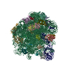
50S ribosomal subunit from Staphylococcus aureus (Strain ATCC43300)
Components
(50S ribosomal protein ...) x 25
23S rRNA
5S rRNA
Keywords
RIBOSOME / 50S subunit / antibiotic resistance
Function / homology
Function and homology information
large ribosomal subunit / transferase activity / 5S rRNA binding / ribosomal large subunit assembly / large ribosomal subunit rRNA binding / cytosolic large ribosomal subunit / cytoplasmic translation / tRNA binding / negative regulation of translation / rRNA binding ...large ribosomal subunit / transferase activity / 5S rRNA binding / ribosomal large subunit assembly / large ribosomal subunit rRNA binding / cytosolic large ribosomal subunit / cytoplasmic translation / tRNA binding / negative regulation of translation / rRNA binding / structural constituent of ribosome / ribosome / translation / ribonucleoprotein complex / mRNA binding / cytoplasm Similarity search - Function
: / Ribosomal Protein L25; Chain P / Ribosomal Protein L25; Chain P / Ribosomal Protein L15; Chain: K; domain 2 - #10 / Ribosomal protein L22/L17 / Ribosomal Protein L15; Chain: K; domain 2 / Ribosomal Protein L15; Chain: K; domain 2 / Ribosomal Protein L22; Chain A / Ribosomal protein L25, long-form / Ribosomal protein L25, beta domain ...: / Ribosomal Protein L25; Chain P / Ribosomal Protein L25; Chain P / Ribosomal Protein L15; Chain: K; domain 2 - #10 / Ribosomal protein L22/L17 / Ribosomal Protein L15; Chain: K; domain 2 / Ribosomal Protein L15; Chain: K; domain 2 / Ribosomal Protein L22; Chain A / Ribosomal protein L25, long-form / Ribosomal protein L25, beta domain / Ribosomal protein L25, C-terminal / Ribosomal protein TL5, C-terminal domain / : / Ribosomal protein L16 signature 1. / Ribosomal protein L16 signature 2. / Ribosomal protein L16, conserved site / : / Ribosomal protein L17 signature. / Ribosomal L25p family / Ribosomal protein L25 / Ribosomal protein L36 signature. / Ribosomal protein L32p, bacterial type / Ribosomal protein L28/L24 superfamily / Ribosomal protein L25/Gln-tRNA synthetase, N-terminal / Ribosomal protein L25/Gln-tRNA synthetase, anti-codon-binding domain superfamily / Ribosomal protein L33, conserved site / Ribosomal protein L33 signature. / Ribosomal protein L35, conserved site / Ribosomal protein L35 signature. / Ribosomal protein L28 / Ribosomal protein L35, non-mitochondrial / Ribosomal protein L18, bacterial-type / : / Ribosomal protein L36 / Ribosomal protein L36 superfamily / Ribosomal protein L36 / Ribosomal protein L19, conserved site / Ribosomal protein L19 signature. / Ribosomal protein L27, conserved site / Ribosomal protein L27 signature. / Ribosomal protein L20 signature. / Ribosomal protein L22, bacterial/chloroplast-type / Ribosomal protein L14P, bacterial-type / Ribosomal protein L34, conserved site / Ribosomal protein L34 signature. / Ribosomal protein L2, bacterial/organellar-type / Ribosomal protein L35 / Ribosomal protein L35 superfamily / Ribosomal protein L35 / Ribosomal protein L33 / Ribosomal protein L18 / Ribosomal L18 of archaea, bacteria, mitoch. and chloroplast / Ribosomal protein L33 / Ribosomal L28 family / Ribosomal protein L33 superfamily / Ribosomal protein L16 / Ribosomal protein L28/L24 / Ribosomal protein L30, bacterial-type / : / L28p-like / Ribosomal protein L27 / Ribosomal L27 protein / Ribosomal protein L20 / Ribosomal L32p protein family / Ribosomal protein L19 / Ribosomal protein L20 / Ribosomal protein L20, C-terminal / Ribosomal protein L19 / Ribosomal protein L19 superfamily / Ribosomal protein L21 / Ribosomal protein L32p / Large ribosomal subunit protein uL24, C-terminal domain / Ribosomal protein L17 / Ribosomal protein L17 superfamily / Ribosomal protein L17 / Ribosomal protein L21-like / L21-like superfamily / Ribosomal prokaryotic L21 protein / Ribosomal protein L34 / Ribosomal protein L34 / Ribosomal protein L24 / Ribosomal protein L3, bacterial/organelle-type / Ribosomal protein L15, bacterial-type / 50S ribosomal protein uL4 / Ribosomal protein L13, bacterial-type / Ribosomal protein L2 signature. / Ribosomal protein L2, conserved site / : / Ribosomal protein L15, conserved site / Ribosomal protein L15 signature. / Ribosomal protein L10e/L16 / Ribosomal protein L10e/L16 superfamily / Ribosomal protein L16p/L10e / Ribosomal protein L13 signature. / Ribosomal protein L13, conserved site / Ribosomal protein L2, domain 3 / Ribosomal protein L22/L17, conserved site / Ribosomal protein L22 signature. / Ribosomal protein L14P, conserved site / Ribosomal protein L14 signature. Similarity search - Domain/homology
: / RNA / RNA (> 10) / RNA (> 100) / RNA (> 1000) / Large ribosomal subunit protein bL17 / Large ribosomal subunit protein uL15 / Large ribosomal subunit protein bL36 / Large ribosomal subunit protein bL28 / Large ribosomal subunit protein uL14 ...: / RNA / RNA (> 10) / RNA (> 100) / RNA (> 1000) / Large ribosomal subunit protein bL17 / Large ribosomal subunit protein uL15 / Large ribosomal subunit protein bL36 / Large ribosomal subunit protein bL28 / Large ribosomal subunit protein uL14 / Large ribosomal subunit protein bL19 / Large ribosomal subunit protein bL32 / Large ribosomal subunit protein bL33 / Large ribosomal subunit protein uL16 / Large ribosomal subunit protein bL20 / Large ribosomal subunit protein uL2 / Large ribosomal subunit protein uL3 / Large ribosomal subunit protein bL35 / Large ribosomal subunit protein bL27 / Large ribosomal subunit protein uL22 / Large ribosomal subunit protein bL25 / 50S ribosomal protein L4 / 50S ribosomal protein L29 / Large ribosomal subunit protein bL21 / Large ribosomal subunit protein bL34 / Large ribosomal subunit protein uL24 / Large ribosomal subunit protein uL18 / Large ribosomal subunit protein uL23 / Large ribosomal subunit protein uL13 / Large ribosomal subunit protein uL30 Similarity search - Component
Biological species
Staphylococcus aureus (bacteria)
Method
ELECTRON MICROSCOPY / single particle reconstruction / cryo EM / Resolution: 3 Å
National Health and Medical Research Council (NHMRC, Australia)
1092262
Australia
Australian Research Council (ARC)
170103567
Australia
Citation
Journal: Microbiol Spectr / Year: 2022 Title: A Structurally Characterized Evolutionary Escape Route from Treatment with the Antibiotic Linezolid. Authors: Laura Perlaza-Jiménez / Kher-Shing Tan / Sarah J Piper / Rachel M Johnson / Rebecca S Bamert / Christopher J Stubenrauch / Alexander Wright / David Lupton / Trevor Lithgow / Matthew J Belousoff / Abstract: Methicillin-resistant Staphylococcus aureus (MRSA) is a bacterial pathogen that presents great health concerns. Treatment requires the use of last-line antibiotics, such as members of the ...Methicillin-resistant Staphylococcus aureus (MRSA) is a bacterial pathogen that presents great health concerns. Treatment requires the use of last-line antibiotics, such as members of the oxazolidinone family, of which linezolid is the first member to see regular use in the clinic. Here, we report a short time scale selection experiment in which strains of MRSA were subjected to linezolid treatment. Clonal isolates which had evolved a linezolid-resistant phenotype were characterized by whole-genome sequencing. Linezolid-resistant mutants were identified which had accumulated mutations in the ribosomal protein uL3. Multiple clones which had two mutations in uL3 exhibited resistance to linezolid, 2-fold higher than the clinical breakpoint. Ribosomes from this strain were isolated and subjected to single-particle cryo-electron microscopic analysis and compared to the ribosomes from the parent strain. We found that the mutations in uL3 lead to a rearrangement of a loop that makes contact with Helix 90, propagating a structural change over 15 Å away. This distal change swings nucleotide U2504 into the binding site of the antibiotic, causing linezolid resistance. Antibiotic resistance poses a critical problem to human health and decreases the utility of these lifesaving drugs. Of particular concern is the "superbug" methicillin-resistant Staphylococcus aureus (MRSA), for which treatment of infection requires the use of last-line antibiotics, including linezolid. In this paper, we characterize the atomic rearrangements which the ribosome, the target of linezolid, undergoes during its evolutionary journey toward becoming drug resistant. Using cryo-electron microscopy, we describe a particular molecular mechanism which MRSA uses to become resistant to linezolid.
History
Deposition
Feb 1, 2022
Deposition site: RCSB / Processing site: RCSB
Revision 1.0
Jul 6, 2022
Provider: repository / Type: Initial release
Revision 1.0
Jul 6, 2022
Data content type: EM metadata / Data content type: EM metadata / Provider: repository / Type: Initial release
Revision 1.0
Jul 6, 2022
Data content type: Additional map / Data content type: Additional map / Provider: repository / Type: Initial release
Revision 1.0
Jul 6, 2022
Data content type: FSC / Data content type: FSC / Provider: repository / Type: Initial release
Revision 1.0
Jul 6, 2022
Data content type: Half map / Part number: 1 / Data content type: Half map / Provider: repository / Type: Initial release
Revision 1.0
Jul 6, 2022
Data content type: Half map / Part number: 2 / Data content type: Half map / Provider: repository / Type: Initial release
Revision 1.0
Jul 6, 2022
Data content type: Image / Data content type: Image / Provider: repository / Type: Initial release
Revision 1.0
Jul 6, 2022
Data content type: Mask / Data content type: Mask / Provider: repository / Type: Initial release
Revision 1.0
Jul 6, 2022
Data content type: Primary map / Data content type: Primary map / Provider: repository / Type: Initial release
Revision 1.0
Jul 6, 2022
Data content type: Additional map / Data content type: Additional map / Provider: repository / Type: Initial release
Revision 1.0
Jul 6, 2022
Data content type: FSC / Data content type: FSC / Provider: repository / Type: Initial release
Revision 1.0
Jul 6, 2022
Data content type: Half map / Part number: 1 / Data content type: Half map / Provider: repository / Type: Initial release
Revision 1.0
Jul 6, 2022
Data content type: Half map / Part number: 2 / Data content type: Half map / Provider: repository / Type: Initial release
Revision 1.0
Jul 6, 2022
Data content type: Image / Data content type: Image / Provider: repository / Type: Initial release
Revision 1.0
Jul 6, 2022
Data content type: Mask / Data content type: Mask / Provider: repository / Type: Initial release
Revision 1.0
Jul 6, 2022
Data content type: Primary map / Data content type: Primary map / Provider: repository / Type: Initial release
Data content type: EM metadata / Data content type: EM metadata / EM metadata / Group: Data processing / Experimental summary / Data content type: EM metadata / EM metadata / Category: em_admin / em_software / Data content type: EM metadata / EM metadata / Item: _em_admin.last_update / _em_software.name
In the structure databanks used in Yorodumi, some data are registered as the other names, "COVID-19 virus" and "2019-nCoV". Here are the details of the virus and the list of structure data.
Jan 31, 2019. EMDB accession codes are about to change! (news from PDBe EMDB page)
EMDB accession codes are about to change! (news from PDBe EMDB page)
The allocation of 4 digits for EMDB accession codes will soon come to an end. Whilst these codes will remain in use, new EMDB accession codes will include an additional digit and will expand incrementally as the available range of codes is exhausted. The current 4-digit format prefixed with “EMD-” (i.e. EMD-XXXX) will advance to a 5-digit format (i.e. EMD-XXXXX), and so on. It is currently estimated that the 4-digit codes will be depleted around Spring 2019, at which point the 5-digit format will come into force.
The EM Navigator/Yorodumi systems omit the EMD- prefix.
Related info.:Q: What is EMD? / ID/Accession-code notation in Yorodumi/EM Navigator
Yorodumi is a browser for structure data from EMDB, PDB, SASBDB, etc.
This page is also the successor to EM Navigator detail page, and also detail information page/front-end page for Omokage search.
The word "yorodu" (or yorozu) is an old Japanese word meaning "ten thousand". "mi" (miru) is to see.
Related info.:EMDB / PDB / SASBDB / Comparison of 3 databanks / Yorodumi Search / Aug 31, 2016. New EM Navigator & Yorodumi / Yorodumi Papers / Jmol/JSmol / Function and homology information / Changes in new EM Navigator and Yorodumi
 Movie
Movie Controller

 Yorodumi
 Open data
 Basic information
 Components
 Keywords
 Function and homology information
 Staphylococcus aureus (bacteria)
 Authors
 United States,
  Australia, 3items
  Citation
 Structure visualization
 Downloads & links
 Other downloads
 PDBj
PDBj


 Assembly
 Sample preparation
 Electron microscopy imaging
 FIELD EMISSION GUN / Accelerating voltage: 200 kV / Illumination mode: FLOOD BEAM
 Processing