Movie
Movie Controller

[English] 日本語
 Yorodumi
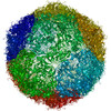
- PDB-7tah: Cryo-EM structure of Human Enterovirus D68 US/MO/14-18947 strain ... -
+ Open data

- Basic information
| Entry | Database: PDB / ID: 7tah | ||||||
|---|---|---|---|---|---|---|---|
| Title | Cryo-EM structure of Human Enterovirus D68 US/MO/14-18947 strain in complex with inhibitor 11526091 (no/low occupancy-no inhibitor modeled) | ||||||
 Components Components |
| ||||||
 Keywords Keywords | VIRUS / EV-D68 / acute flaccid myelitis / AFM / inhibitor / antiviral / Structural Genomics / Center for Structural Genomics of Infectious Diseases / CSGID | ||||||
| Function / homology |  Function and homology information Function and homology informationpicornain 2A / symbiont-mediated suppression of host mRNA export from nucleus / symbiont genome entry into host cell via pore formation in plasma membrane / picornain 3C / T=pseudo3 icosahedral viral capsid / host cell cytoplasmic vesicle membrane / nucleoside-triphosphate phosphatase / channel activity / monoatomic ion transmembrane transport / RNA helicase activity ...picornain 2A / symbiont-mediated suppression of host mRNA export from nucleus / symbiont genome entry into host cell via pore formation in plasma membrane / picornain 3C / T=pseudo3 icosahedral viral capsid / host cell cytoplasmic vesicle membrane / nucleoside-triphosphate phosphatase / channel activity / monoatomic ion transmembrane transport / RNA helicase activity / symbiont-mediated suppression of host innate immune response / endocytosis involved in viral entry into host cell / symbiont-mediated activation of host autophagy / RNA-directed RNA polymerase / cysteine-type endopeptidase activity / viral RNA genome replication / RNA-directed RNA polymerase activity / DNA-templated transcription / virion attachment to host cell / host cell nucleus / structural molecule activity / ATP hydrolysis activity / proteolysis / RNA binding / zinc ion binding / ATP binding / membrane Similarity search - Function | ||||||
| Biological species |  enterovirus D68 enterovirus D68 | ||||||
| Method | ELECTRON MICROSCOPY / single particle reconstruction / cryo EM / Resolution: 2.3 Å | ||||||
 Authors Authors | Fu, J. / Klose, T. / Kuhn, R.J. / Center for Structural Genomics of Infectious Diseases (CSGID) | ||||||
| Funding support |  United States, 1items United States, 1items
| ||||||
 Citation Citation | Journal: To Be Published Title: Isoxazole-3-Carboxamide Derivatives of Pleconaril Destabilize the Viral Capsid of Enterovirus-D68 Authors: Lane, T. / Fu, J. / Sherry, B. / Tarbet, B. / Hurst, B.L. / Riabova, O. / Kazakova, E. / Egorova, A. / Clarke, P. / Leser, S. / Frost, J. / Rudy, M. / Tyler, K. / Klose, T. / Kuhn, R.J. / Makarov, V. / Ekins, S. | ||||||
| History |
|
- Structure visualization
| Structure viewer | Molecule: MolmilJmol/JSmol |
|---|
- Downloads & links
-Download
| PDBx/mmCIF format | 7tah.cif.gz | 153.9 KB | Display | PDBx/mmCIF format |
|---|---|---|---|---|
| PDB format | pdb7tah.ent.gz | 119.5 KB | Display | PDB format |
| PDBx/mmJSON format | 7tah.json.gz | Tree view | PDBx/mmJSON format | |
| Others |  Other downloads Other downloads |
-Validation report
| Summary document | 7tah_validation.pdf.gz | 1.5 MB | Display | wwPDB validaton report |
|---|---|---|---|---|
| Full document | 7tah_full_validation.pdf.gz | 1.5 MB | Display | |
| Data in XML | 7tah_validation.xml.gz | 45 KB | Display | |
| Data in CIF | 7tah_validation.cif.gz | 65.2 KB | Display | |
| Arichive directory | https://data.pdbj.org/pub/pdb/validation_reports/ta/7tahftp://data.pdbj.org/pub/pdb/validation_reports/ta/7tah | HTTPS FTP |
-Related structure data
| Related structure data |  25774MC 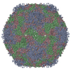 7t9pC 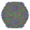 7tafC  7tagC  7tajC M: map data used to model this data C: citing same article ( |
|---|---|
| Similar structure data |

-Links
 PDBj
PDBj

- Assembly
| Deposited unit | 
|
|---|---|
| 1 | x 60
|
| 2 |
|
| 3 | x 5
|
| 4 | x 6
|
| 5 | 
|
| Symmetry | Point symmetry: (Schoenflies symbol: I (icosahedral)) |
-Components
| #1: Protein | Mass: 32819.207 Da / Num. of mol.: 1 / Source method: isolated from a natural source / Source: (natural) enterovirus D68 / Strain: US/MO/14-18947 / References: UniProt: A0A097BW12 |
|---|---|
| #2: Protein | Mass: 27112.814 Da / Num. of mol.: 1 / Fragment: UNP residues 318-564 / Source method: isolated from a natural source / Source: (natural) enterovirus D68 / Strain: US/MO/14-18947 / References: UniProt: A0A097BW12 |
| #3: Protein | Mass: 26573.094 Da / Num. of mol.: 1 / Source method: isolated from a natural source / Source: (natural) enterovirus D68 / Strain: US/MO/14-18947 / References: UniProt: A0A097BW12 |
| #4: Protein | Mass: 7336.960 Da / Num. of mol.: 1 / Fragment: UNP residues 2-69 / Source method: isolated from a natural source / Source: (natural) enterovirus D68 / Strain: US/MO/14-18947 / References: UniProt: A0A097BW12 |
-Experimental details
-Experiment
| Experiment | Method: ELECTRON MICROSCOPY |
|---|---|
| EM experiment | Aggregation state: PARTICLE / 3D reconstruction method: single particle reconstruction |
- Sample preparation
| Component | Name: enterovirus D68 / Type: VIRUS / Entity ID: all / Source: NATURAL |
|---|---|
| Source (natural) | Organism: enterovirus D68 / Strain: US/MO/14-18947 |
| Details of virus | Empty: NO / Enveloped: NO / Isolate: STRAIN / Type: VIRION |
| Natural host | Organism: Homo sapiens |
| Buffer solution | pH: 8 |
| Specimen | Embedding applied: NO / Shadowing applied: NO / Staining applied: NO / Vitrification applied: YES |
| Specimen support | Grid material: COPPER / Grid mesh size: 400 divisions/in. / Grid type: PELCO Ultrathin Carbon with Lacey Carbon |
| Vitrification | Instrument: GATAN CRYOPLUNGE 3 / Cryogen name: ETHANE |
- Electron microscopy imaging
| Experimental equipment |  Model: Titan Krios / Image courtesy: FEI Company |
|---|---|
| Microscopy | Model: FEI TITAN KRIOS |
| Electron gun | Electron source:  FIELD EMISSION GUN / Accelerating voltage: 300 kV / Illumination mode: FLOOD BEAM FIELD EMISSION GUN / Accelerating voltage: 300 kV / Illumination mode: FLOOD BEAM |
| Electron lens | Mode: BRIGHT FIELD / Nominal magnification: 64000 X / Nominal defocus max: 2000 nm / Nominal defocus min: 400 nm / Cs: 2.7 mm / Alignment procedure: COMA FREE |
| Specimen holder | Cryogen: NITROGEN / Specimen holder model: FEI TITAN KRIOS AUTOGRID HOLDER |
| Image recording | Electron dose: 36.06 e/Å2 / Film or detector model: GATAN K3 (6k x 4k) |
| EM imaging optics | Energyfilter name: GIF Bioquantum / Energyfilter slit width: 20 eV |
- Processing
| Software | Name: PHENIX / Version: 1.19.2_4158: / Classification: refinement | ||||||||||||||||||||||||
|---|---|---|---|---|---|---|---|---|---|---|---|---|---|---|---|---|---|---|---|---|---|---|---|---|---|
| EM software |
| ||||||||||||||||||||||||
| CTF correction | Type: PHASE FLIPPING AND AMPLITUDE CORRECTION | ||||||||||||||||||||||||
| Symmetry | Point symmetry: I (icosahedral) | ||||||||||||||||||||||||
| 3D reconstruction | Resolution: 2.3 Å / Resolution method: FSC 0.143 CUT-OFF / Num. of particles: 20971 / Symmetry type: POINT | ||||||||||||||||||||||||
| Atomic model building | Protocol: RIGID BODY FIT / Space: REAL | ||||||||||||||||||||||||
| Atomic model building | PDB-ID: 6CSG Accession code: 6CSG / Source name: PDB / Type: experimental model | ||||||||||||||||||||||||
| Refine LS restraints |
|