 Movie
Movie Controller


+ Open data

- Basic information
| Entry | Database: PDB / ID: 7t5p | |||||||||
|---|---|---|---|---|---|---|---|---|---|---|
| Title | Cryo-EM structure of human SIMC1-SLF2 complex | |||||||||
 Components Components |
| |||||||||
 Keywords Keywords | ANTIVIRAL PROTEIN / Nse5 / Nse6 / SMC5 / SMC6 / SIMC1 / SLF2 / C5orf25 / viral restriction | |||||||||
| Function / homology |  Function and homology information Function and homology informationSUMO polymer binding / positive regulation of maintenance of mitotic sister chromatid cohesion / Smc5-Smc6 complex / peptidase inhibitor activity / chromatin looping / protein localization to site of double-strand break / regulation of telomere maintenance / positive regulation of double-strand break repair / protein sumoylation / sarcomere ...SUMO polymer binding / positive regulation of maintenance of mitotic sister chromatid cohesion / Smc5-Smc6 complex / peptidase inhibitor activity / chromatin looping / protein localization to site of double-strand break / regulation of telomere maintenance / positive regulation of double-strand break repair / protein sumoylation / sarcomere / positive regulation of protein-containing complex assembly / PML body / double-strand break repair via homologous recombination / site of double-strand break / chromosome, telomeric region / DNA damage response / ubiquitin protein ligase binding / chromatin / protein-containing complex binding / nucleoplasm / nucleus / cytoplasm Similarity search - Function | |||||||||
| Biological species |  Homo sapiens (human) Homo sapiens (human) | |||||||||
| Method | ELECTRON MICROSCOPY / single particle reconstruction / cryo EM / Resolution: 3.4 Å | |||||||||
 Authors Authors | Maeda, S. / Oravcova, M. / Boddy, M.N. / Otomo, T. | |||||||||
| Funding support |  United States, 2items United States, 2items
| |||||||||
 Citation Citation | Journal: Elife / Year: 2022 Title: The Nse5/6-like SIMC1-SLF2 complex localizes SMC5/6 to viral replication centers. Authors: Martina Oravcová / Minghua Nie / Nicola Zilio / Shintaro Maeda / Yasaman Jami-Alahmadi / Eros Lazzerini-Denchi / James A Wohlschlegel / Helle D Ulrich / Takanori Otomo / Michael N Boddy /  Abstract: The human SMC5/6 complex is a conserved guardian of genome stability and an emerging component of antiviral responses. These disparate functions likely require distinct mechanisms of SMC5/6 ...The human SMC5/6 complex is a conserved guardian of genome stability and an emerging component of antiviral responses. These disparate functions likely require distinct mechanisms of SMC5/6 regulation. In yeast, Smc5/6 is regulated by its Nse5/6 subunits, but such regulatory subunits for human SMC5/6 are poorly defined. Here, we identify a novel SMC5/6 subunit called SIMC1 that contains SUMO interacting motifs (SIMs) and an Nse5-like domain. We isolated SIMC1 from the proteomic environment of SMC5/6 within polyomavirus large T antigen (LT)-induced subnuclear compartments. SIMC1 uses its SIMs and Nse5-like domain to localize SMC5/6 to polyomavirus replication centers (PyVRCs) at SUMO-rich PML nuclear bodies. SIMC1's Nse5-like domain binds to the putative Nse6 orthologue SLF2 to form an anti-parallel helical dimer resembling the yeast Nse5/6 structure. SIMC1-SLF2 structure-based mutagenesis defines a conserved surface region containing the N-terminus of SIMC1's helical domain that regulates SMC5/6 localization to PyVRCs. Furthermore, SLF1, which recruits SMC5/6 to DNA lesions via its BRCT and ARD motifs, binds SLF2 analogously to SIMC1 and forms a separate Nse5/6-like complex. Thus, two Nse5/6-like complexes with distinct recruitment domains control human SMC5/6 localization. | |||||||||
| History |
|
- Structure visualization
| Structure viewer | Molecule: MolmilJmol/JSmol |
|---|
- Downloads & links
-Download
| PDBx/mmCIF format | 7t5p.cif.gz | 295.5 KB | Display | PDBx/mmCIF format |
|---|---|---|---|---|
| PDB format | pdb7t5p.ent.gz | 235.4 KB | Display | PDB format |
| PDBx/mmJSON format | 7t5p.json.gz | Tree view | PDBx/mmJSON format | |
| Others |  Other downloads Other downloads |
-Validation report
| Arichive directory | https://data.pdbj.org/pub/pdb/validation_reports/t5/7t5pftp://data.pdbj.org/pub/pdb/validation_reports/t5/7t5p | HTTPS FTP |
|---|
-Related structure data
| Related structure data |  25706MC M: map data used to model this data C: citing same article ( |
|---|---|
| Similar structure data |
-Links
 PDBj
PDBj- Assembly
| Deposited unit | 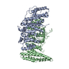
|
|---|---|
| 1 |
|
-Components
| #1: Protein | Mass: 67021.539 Da / Num. of mol.: 1 Source method: isolated from a genetically manipulated source Source: (gene. exp.) Homo sapiens (human) / Gene: SIMC1, C5orf25, PLEIAD / Production host:   Spodoptera frugiperda (fall armyworm) / References: UniProt: Q8NDZ2 Spodoptera frugiperda (fall armyworm) / References: UniProt: Q8NDZ2 |
|---|---|
| #2: Protein | Mass: 61867.793 Da / Num. of mol.: 1 Source method: isolated from a genetically manipulated source Source: (gene. exp.) Homo sapiens (human) / Gene: SLF2, C10orf6, FAM178A / Production host: Spodoptera frugiperda (fall armyworm) / References: UniProt: Q8IX21 |
-Experimental details
-Experiment
| Experiment | Method: ELECTRON MICROSCOPY |
|---|---|
| EM experiment | Aggregation state: PARTICLE / 3D reconstruction method: single particle reconstruction |
- Sample preparation
| Component | Name: Human SIMC1-SLF2 complex / Type: COMPLEX / Entity ID: all / Source: RECOMBINANT |
|---|---|
| Molecular weight | Experimental value: NO |
| Source (natural) | Organism: Homo sapiens (human) |
| Source (recombinant) | Organism: Spodoptera frugiperda (fall armyworm) |
| Buffer solution | pH: 7.5 |
| Specimen | Conc.: 0.4 mg/ml / Embedding applied: NO / Shadowing applied: NO / Staining applied: NO / Vitrification applied: YES |
| Vitrification | Instrument: HOMEMADE PLUNGER / Cryogen name: ETHANE / Humidity: 88 % / Chamber temperature: 277 K |
- Electron microscopy imaging
| Experimental equipment |  Model: Talos Arctica / Image courtesy: FEI Company |
|---|---|
| Microscopy | Model: FEI TALOS ARCTICA |
| Electron gun | Electron source:  FIELD EMISSION GUN / Accelerating voltage: 200 kV / Illumination mode: FLOOD BEAM FIELD EMISSION GUN / Accelerating voltage: 200 kV / Illumination mode: FLOOD BEAM |
| Electron lens | Mode: BRIGHT FIELD / Nominal magnification: 73000 X / Nominal defocus max: 1800 nm / Nominal defocus min: 500 nm / Cs: 2.7 mm / C2 aperture diameter: 70 µm / Alignment procedure: COMA FREE |
| Specimen holder | Cryogen: NITROGEN / Specimen holder model: FEI TITAN KRIOS AUTOGRID HOLDER |
| Image recording | Electron dose: 66 e/Å2 / Detector mode: COUNTING / Film or detector model: GATAN K2 SUMMIT (4k x 4k) |
- Processing
| EM software |
| |||||||||||||||||||||||||||
|---|---|---|---|---|---|---|---|---|---|---|---|---|---|---|---|---|---|---|---|---|---|---|---|---|---|---|---|---|
| CTF correction | Type: PHASE FLIPPING AND AMPLITUDE CORRECTION | |||||||||||||||||||||||||||
| 3D reconstruction | Resolution: 3.4 Å / Resolution method: FSC 0.143 CUT-OFF / Num. of particles: 39826 / Symmetry type: POINT | |||||||||||||||||||||||||||
| Atomic model building | Protocol: RIGID BODY FIT / Space: REAL |
