 Movie
Movie Controller

[English] 日本語
 Yorodumi
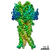
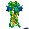
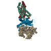
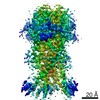
- PDB-7sqh: Structure of the human proton-activated chloride channel ASOR in ... -
+ Open data

- Basic information
| Entry | Database: PDB / ID: 7sqh | |||||||||
|---|---|---|---|---|---|---|---|---|---|---|
| Title | Structure of the human proton-activated chloride channel ASOR in desensitized conformation | |||||||||
 Components Components | Proton-activated chloride channel | |||||||||
 Keywords Keywords | MEMBRANE PROTEIN / ion channel / chloride channel | |||||||||
| Function / homology | pH-gated chloride channel activity / TMEM206 protein / TMEM206 protein family / chloride transport / chloride channel complex / cell surface / plasma membrane / Proton-activated chloride channel Function and homology information Function and homology information | |||||||||
| Biological species |  Homo sapiens (human) Homo sapiens (human) | |||||||||
| Method | ELECTRON MICROSCOPY / single particle reconstruction / cryo EM / Resolution: 2.5 Å | |||||||||
 Authors Authors | Long, S.B. / Wang, C. / Delgado, B. | |||||||||
| Funding support |  United States, 2items United States, 2items
| |||||||||
 Citation Citation | Journal: Sci Adv / Year: 2022 Title: Gating choreography and mechanism of the human proton-activated chloride channel ASOR. Authors: Chongyuan Wang / Maya M Polovitskaya / Bryce D Delgado / Thomas J Jentsch / Stephen B Long /  Abstract: The proton-activated chloride channel ASOR (TMEM206/PAC) permeates anions across cellular membranes in response to acidification, thereby enhancing acid-induced cell death and regulating endocytosis. ...The proton-activated chloride channel ASOR (TMEM206/PAC) permeates anions across cellular membranes in response to acidification, thereby enhancing acid-induced cell death and regulating endocytosis. The molecular mechanisms of pH-dependent control are not understood, in part because structural information for an activated conformation of ASOR is lacking. Here, we reconstitute function from purified protein and present a 3.1-Å-resolution cryo-electron microscopy structure of human ASOR at acidic pH in an activated conformation. The work contextualizes a previous acidic pH structure as a desensitized conformation. Combined with electrophysiological studies and high-resolution structures of resting and desensitized states, the work reveals mechanisms of proton sensing and ion pore gating. Clusters of extracellular acidic residues function as pH sensors and coalesce when protonated. Ensuing conformational changes induce metamorphosis of transmembrane helices to fashion an ion conduction pathway unique to the activated conformation. The studies identify a new paradigm of channel gating in this ubiquitous ion channel. | |||||||||
| History |
|
- Structure visualization
| Movie |
Movie viewer |
|---|---|
| Structure viewer | Molecule: MolmilJmol/JSmol |
- Downloads & links
-Download
| PDBx/mmCIF format | 7sqh.cif.gz | 164.5 KB | Display | PDBx/mmCIF format |
|---|---|---|---|---|
| PDB format | pdb7sqh.ent.gz | Display | PDB format | |
| PDBx/mmJSON format | 7sqh.json.gz | Tree view | PDBx/mmJSON format | |
| Others |  Other downloads Other downloads |
-Validation report
| Arichive directory | https://data.pdbj.org/pub/pdb/validation_reports/sq/7sqhftp://data.pdbj.org/pub/pdb/validation_reports/sq/7sqh | HTTPS FTP |
|---|
-Related structure data
| Related structure data | 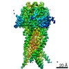 25385MC  7sqfC  7sqgC C: citing same article ( M: map data used to model this data |
|---|---|
| Similar structure data |


-Links
 PDBj
PDBj- Assembly
| Deposited unit | 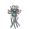
|
|---|---|
| 1 |
|
-Components
| #1: Protein | Mass: 40092.047 Da / Num. of mol.: 3 Source method: isolated from a genetically manipulated source Source: (gene. exp.) Homo sapiens (human) / Gene: PACC1, C1orf75, TMEM206 / Production host: Homo sapiens (human) / References: UniProt: Q9H813#2: Sugar | ChemComp-NAG /   Type: D-saccharide, beta linking / Mass: 221.208 Da / Num. of mol.: 12 Type: D-saccharide, beta linking / Mass: 221.208 Da / Num. of mol.: 12Source method: isolated from a genetically manipulated source Formula: C8H15NO6 #3: Water | ChemComp-HOH / |  Mass: 18.015 Da / Num. of mol.: 21 / Source method: isolated from a natural source / Formula: H2O Mass: 18.015 Da / Num. of mol.: 21 / Source method: isolated from a natural source / Formula: H2OHas ligand of interest | N | Has protein modification | Y | |
|---|
-Experimental details
-Experiment
| Experiment | Method: ELECTRON MICROSCOPY |
|---|---|
| EM experiment | Aggregation state: PARTICLE / 3D reconstruction method: single particle reconstruction |
- Sample preparation
| Component | Name: proton activated chloride channel TMEM206 in resting conformation Type: COMPLEX / Entity ID: #1 / Source: RECOMBINANT | ||||||||||||||||||||
|---|---|---|---|---|---|---|---|---|---|---|---|---|---|---|---|---|---|---|---|---|---|
| Molecular weight | Units: MEGADALTONS / Experimental value: NO | ||||||||||||||||||||
| Source (natural) | Organism: Homo sapiens (human) | ||||||||||||||||||||
| Source (recombinant) | Organism: Homo sapiens (human) / Cell: Expi293, Invitrogen | ||||||||||||||||||||
| Buffer solution | pH: 4.5 | ||||||||||||||||||||
| Buffer component |
| ||||||||||||||||||||
| Specimen | Conc.: 1.6 mg/ml / Embedding applied: NO / Shadowing applied: NO / Staining applied: NO / Vitrification applied: YES / Details: Monodisperse sample | ||||||||||||||||||||
| Specimen support | Grid type: UltrAuFoil R1.2/1.3 | ||||||||||||||||||||
| Vitrification | Instrument: FEI VITROBOT MARK IV / Cryogen name: ETHANE / Humidity: 100 % / Chamber temperature: 298 K / Details: 2 second blot, blot force of 0 |
- Electron microscopy imaging
| Experimental equipment |  Model: Titan Krios / Image courtesy: FEI Company |
|---|---|
| Microscopy | Model: FEI TITAN KRIOS |
| Electron gun | Electron source:  FIELD EMISSION GUN / Accelerating voltage: 300 kV / Illumination mode: FLOOD BEAM FIELD EMISSION GUN / Accelerating voltage: 300 kV / Illumination mode: FLOOD BEAM |
| Electron lens | Mode: BRIGHT FIELD / Nominal magnification: 22500 X / Nominal defocus max: -3000 nm / Nominal defocus min: -1000 nm / Alignment procedure: BASIC |
| Specimen holder | Cryogen: NITROGEN / Specimen holder model: FEI TITAN KRIOS AUTOGRID HOLDER |
| Image recording | Average exposure time: 4 sec. / Electron dose: 71 e/Å2 / Detector mode: SUPER-RESOLUTION / Film or detector model: GATAN K3 BIOQUANTUM (6k x 4k) / Num. of grids imaged: 1 / Num. of real images: 157906 |
| Image scans | Movie frames/image: 40 / Used frames/image: 1-40 |
- Processing
| Software | Name: PHENIX / Version: 1.19.1_4122: / Classification: refinement | ||||||||||||||||||||||||||||||||||||||||
|---|---|---|---|---|---|---|---|---|---|---|---|---|---|---|---|---|---|---|---|---|---|---|---|---|---|---|---|---|---|---|---|---|---|---|---|---|---|---|---|---|---|
| EM software |
| ||||||||||||||||||||||||||||||||||||||||
| CTF correction | Type: PHASE FLIPPING AND AMPLITUDE CORRECTION | ||||||||||||||||||||||||||||||||||||||||
| Particle selection | Num. of particles selected: 3097510 | ||||||||||||||||||||||||||||||||||||||||
| 3D reconstruction | Resolution: 2.5 Å / Resolution method: FSC 0.143 CUT-OFF / Num. of particles: 157906 / Symmetry type: POINT | ||||||||||||||||||||||||||||||||||||||||
| Atomic model building | B value: 30 / Protocol: AB INITIO MODEL / Space: REAL / Target criteria: Correlation coefficient | ||||||||||||||||||||||||||||||||||||||||
| Refine LS restraints |
|
