[English] 日本語
 Yorodumi
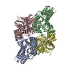
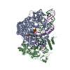
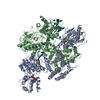
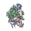
- PDB-7sjr: Cryo-EM structure of AdnA-AdnB(W325A) in complex with DNA and AMPPNP -
+ Open data
- Basic information
| Entry | Database: PDB / ID: 7sjr | |||||||||||||||||||||||||||||||||
|---|---|---|---|---|---|---|---|---|---|---|---|---|---|---|---|---|---|---|---|---|---|---|---|---|---|---|---|---|---|---|---|---|---|---|
| Title | Cryo-EM structure of AdnA-AdnB(W325A) in complex with DNA and AMPPNP | |||||||||||||||||||||||||||||||||
 Components Components |
| |||||||||||||||||||||||||||||||||
 Keywords Keywords | DNA BINDING PROTEIN/DNA / homologous recombination / DNA end resection / cryoelectron microscopy / DNA curtain / DNA BINDING PROTEIN / DNA BINDING PROTEIN-DNA complex | |||||||||||||||||||||||||||||||||
| Function / homology |  Function and homology information Function and homology informationDNA helicase complex / recombinational repair / exonuclease activity / DNA 3'-5' helicase / 3'-5' DNA helicase activity / DNA helicase activity / DNA helicase / hydrolase activity / DNA repair / DNA binding ...DNA helicase complex / recombinational repair / exonuclease activity / DNA 3'-5' helicase / 3'-5' DNA helicase activity / DNA helicase activity / DNA helicase / hydrolase activity / DNA repair / DNA binding / ATP binding / cytosol Similarity search - Function | |||||||||||||||||||||||||||||||||
| Biological species |  Mycolicibacterium smegmatis (bacteria) Mycolicibacterium smegmatis (bacteria) | |||||||||||||||||||||||||||||||||
| Method | ELECTRON MICROSCOPY / single particle reconstruction / cryo EM / Resolution: 3.8 Å | |||||||||||||||||||||||||||||||||
 Authors Authors | Wang, J. / Warren, G.M. / Shuman, S. / Patel, D.J. | |||||||||||||||||||||||||||||||||
| Funding support |  United States, 2items United States, 2items
| |||||||||||||||||||||||||||||||||
 Citation Citation |  Journal: Nucleic Acids Res / Year: 2022 Journal: Nucleic Acids Res / Year: 2022Title: Structure-activity relationships at a nucleobase-stacking tryptophan required for chemomechanical coupling in the DNA resecting motor-nuclease AdnAB. Authors: Garrett M Warren / Aviv Meir / Juncheng Wang / Dinshaw J Patel / Eric C Greene / Stewart Shuman /  Abstract: Mycobacterial AdnAB is a heterodimeric helicase-nuclease that initiates homologous recombination by resecting DNA double-strand breaks. The AdnB subunit hydrolyzes ATP to drive single-nucleotide ...Mycobacterial AdnAB is a heterodimeric helicase-nuclease that initiates homologous recombination by resecting DNA double-strand breaks. The AdnB subunit hydrolyzes ATP to drive single-nucleotide steps of 3'-to-5' translocation of AdnAB on the tracking DNA strand via a ratchet-like mechanism. Trp325 in AdnB motif III, which intercalates into the tracking strand and makes a π stack on a nucleobase 5' of a flipped-out nucleoside, is the putative ratchet pawl without which ATP hydrolysis is mechanically futile. Here, we report that AdnAB mutants wherein Trp325 was replaced with phenylalanine, tyrosine, histidine, leucine, or alanine retained activity in ssDNA-dependent ATP hydrolysis but displayed a gradient of effects on DSB resection. The resection velocities of Phe325 and Tyr325 mutants were 90% and 85% of the wild-type AdnAB velocity. His325 slowed resection rate to 3% of wild-type and Leu325 and Ala325 abolished DNA resection. A cryo-EM structure of the DNA-bound Ala325 mutant revealed that the AdnB motif III peptide was disordered and the erstwhile flipped out tracking strand nucleobase reverted to a continuous base-stacked arrangement with its neighbors. We conclude that π stacking of Trp325 on a DNA nucleobase triggers and stabilizes the flipped-out conformation of the neighboring nucleoside that underlies formation of a ratchet pawl. | |||||||||||||||||||||||||||||||||
| History |
|
- Structure visualization
| Movie |
 Movie viewer Movie viewer |
|---|---|
| Structure viewer | Molecule:  Molmil Molmil Jmol/JSmol Jmol/JSmol |
- Downloads & links
- Download
| PDBx/mmCIF format |  7sjr.cif.gz 7sjr.cif.gz | 328.8 KB | Display |  PDBx/mmCIF format PDBx/mmCIF format |
|---|---|---|---|---|
| PDB format |  pdb7sjr.ent.gz pdb7sjr.ent.gz | 250.2 KB | Display |  PDB format PDB format |
| PDBx/mmJSON format |  7sjr.json.gz 7sjr.json.gz | Tree view |  PDBx/mmJSON format PDBx/mmJSON format | |
| Others |  Other downloads Other downloads |
-Validation report
| Summary document |  7sjr_validation.pdf.gz 7sjr_validation.pdf.gz | 1010.6 KB | Display |  wwPDB validaton report wwPDB validaton report |
|---|---|---|---|---|
| Full document |  7sjr_full_validation.pdf.gz 7sjr_full_validation.pdf.gz | 1 MB | Display | |
| Data in XML |  7sjr_validation.xml.gz 7sjr_validation.xml.gz | 49.2 KB | Display | |
| Data in CIF |  7sjr_validation.cif.gz 7sjr_validation.cif.gz | 77.2 KB | Display | |
| Arichive directory |  https://data.pdbj.org/pub/pdb/validation_reports/sj/7sjr https://data.pdbj.org/pub/pdb/validation_reports/sj/7sjr ftp://data.pdbj.org/pub/pdb/validation_reports/sj/7sjr ftp://data.pdbj.org/pub/pdb/validation_reports/sj/7sjr | HTTPS FTP |
-Related structure data
| Related structure data |  25164MC M: map data used to model this data C: citing same article ( |
|---|---|
| Similar structure data |
- Links
Links
- Assembly
| Deposited unit | 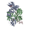
|
|---|---|
| 1 |
|
- Components
-Protein , 2 types, 2 molecules BA
| #1: Protein | Mass: 118013.406 Da / Num. of mol.: 1 Source method: isolated from a genetically manipulated source Source: (gene. exp.)  Mycolicibacterium smegmatis (bacteria) / Gene: pcrA, BIN_B_03433 / Production host: Mycolicibacterium smegmatis (bacteria) / Gene: pcrA, BIN_B_03433 / Production host:  |
|---|---|
| #2: Protein | Mass: 111030.656 Da / Num. of mol.: 1 Source method: isolated from a genetically manipulated source Source: (gene. exp.)  Mycolicibacterium smegmatis (bacteria) / Gene: pcrA_1, ERS451418_01973 / Production host: Mycolicibacterium smegmatis (bacteria) / Gene: pcrA_1, ERS451418_01973 / Production host:  |
-DNA chain , 1 types, 1 molecules C
| #3: DNA chain | Mass: 21477.703 Da / Num. of mol.: 1 / Source method: obtained synthetically / Source: (synth.)  Mycolicibacterium smegmatis (bacteria) Mycolicibacterium smegmatis (bacteria) |
|---|
-Non-polymers , 3 types, 3 molecules 


| #4: Chemical | ChemComp-MG / |
|---|---|
| #5: Chemical | ChemComp-SF4 / |
| #6: Chemical | ChemComp-ANP / |
-Details
| Has ligand of interest | Y |
|---|---|
| Has protein modification | N |
-Experimental details
-Experiment
| Experiment | Method: ELECTRON MICROSCOPY |
|---|---|
| EM experiment | Aggregation state: PARTICLE / 3D reconstruction method: single particle reconstruction |
- Sample preparation
| Component | Name: Cryo-EM map of AdnA-AdnB(W325A) in complex with DNA and AMPPNP Type: COMPLEX / Entity ID: #1-#3 / Source: RECOMBINANT |
|---|---|
| Molecular weight | Experimental value: NO |
| Source (natural) | Organism:  Mycolicibacterium smegmatis (bacteria) Mycolicibacterium smegmatis (bacteria) |
| Source (recombinant) | Organism:  |
| Buffer solution | pH: 8 |
| Specimen | Embedding applied: NO / Shadowing applied: NO / Staining applied: NO / Vitrification applied: YES |
| Specimen support | Grid material: GOLD / Grid type: UltrAuFoil R1.2/1.3 |
| Vitrification | Cryogen name: ETHANE / Humidity: 100 % |
- Electron microscopy imaging
| Experimental equipment |  Model: Titan Krios / Image courtesy: FEI Company |
|---|---|
| Microscopy | Model: FEI TITAN KRIOS |
| Electron gun | Electron source:  FIELD EMISSION GUN / Accelerating voltage: 300 kV / Illumination mode: FLOOD BEAM FIELD EMISSION GUN / Accelerating voltage: 300 kV / Illumination mode: FLOOD BEAM |
| Electron lens | Mode: BRIGHT FIELD / Alignment procedure: COMA FREE |
| Specimen holder | Cryogen: NITROGEN / Specimen holder model: FEI TITAN KRIOS AUTOGRID HOLDER |
| Image recording | Electron dose: 53 e/Å2 / Film or detector model: GATAN K3 (6k x 4k) |
- Processing
| Software | Name: PHENIX / Version: 1.18.2_3874: / Classification: refinement | ||||||||||||||||||||||||
|---|---|---|---|---|---|---|---|---|---|---|---|---|---|---|---|---|---|---|---|---|---|---|---|---|---|
| EM software | Name: PHENIX / Category: model refinement | ||||||||||||||||||||||||
| CTF correction | Type: PHASE FLIPPING AND AMPLITUDE CORRECTION | ||||||||||||||||||||||||
| 3D reconstruction | Resolution: 3.8 Å / Resolution method: FSC 0.143 CUT-OFF / Num. of particles: 98408 / Symmetry type: POINT | ||||||||||||||||||||||||
| Atomic model building | Protocol: RIGID BODY FIT / Space: REAL | ||||||||||||||||||||||||
| Atomic model building | PDB-ID: 6PPJ Accession code: 6PPJ / Source name: PDB / Type: experimental model | ||||||||||||||||||||||||
| Refine LS restraints |
|
 Movie
Movie Controller


 PDBj
PDBj


