 Movie
Movie Controller


+ Open data

- Basic information
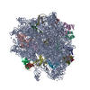
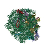
| Entry | Database: PDB / ID: 7s9u | ||||||
|---|---|---|---|---|---|---|---|
| Title | 44SR3C ribosomal particle | ||||||
 Components Components |
| ||||||
 Keywords Keywords | RIBOSOME / Ribosome assembly / 50S subunit / RbgA / YlqF / uL6 | ||||||
| Function / homology |  Function and homology information Function and homology informationlarge ribosomal subunit / transferase activity / large ribosomal subunit rRNA binding / cytosolic large ribosomal subunit / cytoplasmic translation / rRNA binding / negative regulation of translation / ribosome / structural constituent of ribosome / ribonucleoprotein complex ...large ribosomal subunit / transferase activity / large ribosomal subunit rRNA binding / cytosolic large ribosomal subunit / cytoplasmic translation / rRNA binding / negative regulation of translation / ribosome / structural constituent of ribosome / ribonucleoprotein complex / translation / mRNA binding / cytoplasm Similarity search - Function | ||||||
| Biological species |  | ||||||
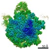
| Method | ELECTRON MICROSCOPY / single particle reconstruction / cryo EM / Resolution: 3.2 Å | ||||||
 Authors Authors | Ortega, J. / Seffouh, A. | ||||||
| Funding support |  Canada, 1items Canada, 1items
| ||||||
 Citation Citation | Journal: Nucleic Acids Res / Year: 2022 Title: RbgA ensures the correct timing in the maturation of the 50S subunits functional sites. Authors: Amal Seffouh / Chirstian Trahan / Tanzila Wasi / Nikhil Jain / Kaustuv Basu / Robert A Britton / Marlene Oeffinger / Joaquin Ortega /  Abstract: RbgA is an essential protein for the assembly of the 50S subunit in Bacillus subtilis. Depletion of RbgA leads to the accumulation of the 45S intermediate. A strain expressing a RbgA variant with ...RbgA is an essential protein for the assembly of the 50S subunit in Bacillus subtilis. Depletion of RbgA leads to the accumulation of the 45S intermediate. A strain expressing a RbgA variant with reduced GTPase activity generates spontaneous suppressor mutations in uL6. Each suppressor strain accumulates a unique 44S intermediate. We reasoned that characterizing the structure of these mutant 44S intermediates may explain why RbgA is required to catalyze the folding of the 50S functional sites. We found that in the 44S particles, rRNA helices H42 and H97, near the binding site of uL6, adopt a flexible conformation and allow the central protuberance and functional sites in the mutant 44S particles to mature in any order. Instead, the wild-type 45S particles exhibit a stable H42-H97 interaction and their functional sites always mature last. The dependence on RbgA was also less pronounced in the 44S particles. We concluded that the binding of uL6 pauses the maturation of the functional sites, but the central protuberance continues to fold. RbgA exclusively binds intermediates with a formed central protuberance and licenses the folding of the functional sites. Through this mechanism, RbgA ensures that the functional sites of the 50S mature last. | ||||||
| History |
|
- Structure visualization
| Movie |
Movie viewer |
|---|---|
| Structure viewer | Molecule: MolmilJmol/JSmol |
- Downloads & links
-Download
| PDBx/mmCIF format | 7s9u.cif.gz | 1.5 MB | Display | PDBx/mmCIF format |
|---|---|---|---|---|
| PDB format | pdb7s9u.ent.gz | 1.1 MB | Display | PDB format |
| PDBx/mmJSON format | 7s9u.json.gz | Tree view | PDBx/mmJSON format | |
| Others |  Other downloads Other downloads |
-Validation report
| Summary document | 7s9u_validation.pdf.gz | 1 MB | Display | wwPDB validaton report |
|---|---|---|---|---|
| Full document | 7s9u_full_validation.pdf.gz | 1.2 MB | Display | |
| Data in XML | 7s9u_validation.xml.gz | 114.8 KB | Display | |
| Data in CIF | 7s9u_validation.cif.gz | 191.5 KB | Display | |
| Arichive directory | https://data.pdbj.org/pub/pdb/validation_reports/s9/7s9uftp://data.pdbj.org/pub/pdb/validation_reports/s9/7s9u | HTTPS FTP |
-Related structure data
| Related structure data |  24937MC  7saeC M: map data used to model this data C: citing same article ( |
|---|---|
| Similar structure data |


-Links
 PDBj
PDBj


- Assembly
| Deposited unit | 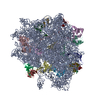
|
|---|---|
| 1 |
|
-Components
-RNA chain , 1 types, 1 molecules A
| #1: RNA chain | Mass: 949606.250 Da / Num. of mol.: 1 / Source method: isolated from a natural source / Source: (natural) |
|---|
-50S ribosomal protein ... , 18 types, 18 molecules CDEJKLNPQRSTUVZbYd
| #2: Protein | Mass: 30335.125 Da / Num. of mol.: 1 / Source method: isolated from a natural source / Source: (natural) |
|---|---|
| #3: Protein | Mass: 22723.348 Da / Num. of mol.: 1 / Source method: isolated from a natural source / Source: (natural) |
| #4: Protein | Mass: 22424.951 Da / Num. of mol.: 1 / Source method: isolated from a natural source / Source: (natural) |
| #5: Protein | Mass: 16407.104 Da / Num. of mol.: 1 / Source method: isolated from a natural source / Source: (natural) |
| #6: Protein | Mass: 13175.288 Da / Num. of mol.: 1 / Source method: isolated from a natural source / Source: (natural) |
| #7: Protein | Mass: 15410.694 Da / Num. of mol.: 1 / Source method: isolated from a natural source / Source: (natural) |
| #8: Protein | Mass: 13774.806 Da / Num. of mol.: 1 / Source method: isolated from a natural source / Source: (natural) |
| #9: Protein | Mass: 13416.853 Da / Num. of mol.: 1 / Source method: isolated from a natural source / Source: (natural) |
| #10: Protein | Mass: 13669.189 Da / Num. of mol.: 1 / Source method: isolated from a natural source / Source: (natural) |
| #11: Protein | Mass: 11296.081 Da / Num. of mol.: 1 / Source method: isolated from a natural source / Source: (natural) |
| #12: Protein | Mass: 12481.608 Da / Num. of mol.: 1 / Source method: isolated from a natural source / Source: (natural) |
| #13: Protein | Mass: 10978.813 Da / Num. of mol.: 1 / Source method: isolated from a natural source / Source: (natural) |
| #14: Protein | Mass: 11166.120 Da / Num. of mol.: 1 / Source method: isolated from a natural source / Source: (natural) |
| #15: Protein | Mass: 10391.855 Da / Num. of mol.: 1 / Source method: isolated from a natural source / Source: (natural) |
| #16: Protein | Mass: 6650.795 Da / Num. of mol.: 1 / Source method: isolated from a natural source / Source: (natural) |
| #17: Protein | Mass: 6745.073 Da / Num. of mol.: 1 / Source method: isolated from a natural source / Source: (natural) |
| #18: Protein | Mass: 7728.029 Da / Num. of mol.: 1 / Source method: isolated from a natural source / Source: (natural) |
| #19: Protein/peptide | Mass: 5271.332 Da / Num. of mol.: 1 / Source method: isolated from a natural source / Source: (natural) |
-Experimental details
-Experiment
| Experiment | Method: ELECTRON MICROSCOPY |
|---|---|
| EM experiment | Aggregation state: PARTICLE / 3D reconstruction method: single particle reconstruction |
- Sample preparation
| Component | Name: 44S_R3C_ribosomal_particle / Type: RIBOSOME / Entity ID: all / Source: NATURAL |
|---|---|
| Source (natural) | Organism: |
| Buffer solution | pH: 7.5 |
| Specimen | Embedding applied: NO / Shadowing applied: NO / Staining applied: NO / Vitrification applied: YES |
| Vitrification | Cryogen name: ETHANE |
- Electron microscopy imaging
| Experimental equipment |  Model: Titan Krios / Image courtesy: FEI Company |
|---|---|
| Microscopy | Model: FEI TITAN KRIOS |
| Electron gun | Electron source:  FIELD EMISSION GUN / Accelerating voltage: 300 kV / Illumination mode: FLOOD BEAM FIELD EMISSION GUN / Accelerating voltage: 300 kV / Illumination mode: FLOOD BEAM |
| Electron lens | Mode: BRIGHT FIELD / Nominal defocus max: 3250 nm / Nominal defocus min: 1250 nm |
| Image recording | Electron dose: 50 e/Å2 / Film or detector model: GATAN K3 BIOQUANTUM (6k x 4k) |
- Processing
| Software | Name: PHENIX / Version: 1.14_3260: / Classification: refinement |
|---|---|
| CTF correction | Type: PHASE FLIPPING AND AMPLITUDE CORRECTION |
| 3D reconstruction | Resolution: 3.2 Å / Resolution method: FSC 0.143 CUT-OFF / Num. of particles: 121252 / Symmetry type: POINT |
