Movie
Movie Controller

[English] 日本語
 Yorodumi
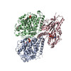
- PDB-7rx0: Complex of AMPPNP-Kif7 and Gli2 Zinc-Finger domain bound to micro... -
+ Open data

- Basic information
| Entry | Database: PDB / ID: 7rx0 | ||||||
|---|---|---|---|---|---|---|---|
| Title | Complex of AMPPNP-Kif7 and Gli2 Zinc-Finger domain bound to microtubules | ||||||
 Components Components |
| ||||||
 Keywords Keywords | MOTOR PROTEIN / Kinesin / Microtubule / Transcription factor / motor domain | ||||||
| Function / homology |  Function and homology information Function and homology informationpositive regulation of smoothened signaling pathway / microtubule motor activity / kinesin complex / microtubule-based movement / Hedgehog 'off' state / ciliary tip / negative regulation of smoothened signaling pathway / Hedgehog 'on' state / structural constituent of cytoskeleton / microtubule cytoskeleton organization ...positive regulation of smoothened signaling pathway / microtubule motor activity / kinesin complex / microtubule-based movement / Hedgehog 'off' state / ciliary tip / negative regulation of smoothened signaling pathway / Hedgehog 'on' state / structural constituent of cytoskeleton / microtubule cytoskeleton organization / neuron migration / fibrillar center / mitotic cell cycle / microtubule binding / microtubule / Hydrolases; Acting on acid anhydrides; Acting on GTP to facilitate cellular and subcellular movement / cilium / ciliary basal body / hydrolase activity / GTPase activity / GTP binding / ATP hydrolysis activity / ATP binding / metal ion binding / cytoplasm / cytosol Similarity search - Function | ||||||
| Biological species |  Homo sapiens (human) Homo sapiens (human) | ||||||
| Method | ELECTRON MICROSCOPY / helical reconstruction / cryo EM / Resolution: 3.89 Å | ||||||
 Authors Authors | Mani, N. / Wilson-Kubalek, E.M. / Haque, F. / Freniere, C. / Milligan, R.A. / Subramanian, R. | ||||||
| Funding support |  United States, 1items United States, 1items
| ||||||
 Citation Citation | Journal: Nat Cell Biol / Year: 2022 Title: Cytoskeletal regulation of a transcription factor by DNA mimicry via coiled-coil interactions. Authors: Farah Haque / Christian Freniere / Qiong Ye / Nandini Mani / Elizabeth M Wilson-Kubalek / Pei-I Ku / Ronald A Milligan / Radhika Subramanian / Abstract: A long-established strategy for transcription regulation is the tethering of transcription factors to cellular membranes. By contrast, the principal effectors of Hedgehog signalling, the GLI ...A long-established strategy for transcription regulation is the tethering of transcription factors to cellular membranes. By contrast, the principal effectors of Hedgehog signalling, the GLI transcription factors, are regulated by microtubules in the primary cilium and the cytoplasm. How GLI is tethered to microtubules remains unclear. Here, we uncover DNA mimicry by the ciliary kinesin KIF7 as a mechanism for the recruitment of GLI to microtubules, wherein the coiled-coil dimerization domain of KIF7, characterized by its striking shape, size and charge similarity to DNA, forms a complex with the DNA-binding zinc fingers in GLI, thus revealing a mode of tethering a DNA-binding protein to the cytoskeleton. GLI increases KIF7 microtubule affinity and consequently modulates the localization of both proteins to microtubules and the cilium tip. Thus, the kinesin-microtubule system is not a passive GLI tether but a regulatable platform tuned by the kinesin-transcription factor interaction. We retooled this coiled-coil-based GLI-KIF7 interaction to inhibit the nuclear and cilium localization of GLI. This strategy can potentially be exploited to downregulate erroneously activated GLI in human cancers. | ||||||
| History |
|
- Structure visualization
| Structure viewer | Molecule: MolmilJmol/JSmol |
|---|
- Downloads & links
-Download
| PDBx/mmCIF format | 7rx0.cif.gz | 237.5 KB | Display | PDBx/mmCIF format |
|---|---|---|---|---|
| PDB format | pdb7rx0.ent.gz | 180.4 KB | Display | PDB format |
| PDBx/mmJSON format | 7rx0.json.gz | Tree view | PDBx/mmJSON format | |
| Others |  Other downloads Other downloads |
-Validation report
| Arichive directory | https://data.pdbj.org/pub/pdb/validation_reports/rx/7rx0ftp://data.pdbj.org/pub/pdb/validation_reports/rx/7rx0 | HTTPS FTP |
|---|
-Related structure data
| Related structure data |  24721MC M: map data used to model this data C: citing same article ( |
|---|---|
| Similar structure data |
-Links
 PDBj
PDBj


- Assembly
| Deposited unit | 
|
|---|---|
| 1 |
|
-Components
-Protein , 4 types, 4 molecules CGAB
| #1: Protein | Mass: 59525.902 Da / Num. of mol.: 1 Source method: isolated from a genetically manipulated source Source: (gene. exp.) Homo sapiens (human) / Gene: KIF7, UNQ340/PRO539 / Production host:   Spodoptera frugiperda (fall armyworm) / References: UniProt: Q2M1P5 Spodoptera frugiperda (fall armyworm) / References: UniProt: Q2M1P5 |
|---|---|
| #2: Protein | Mass: 7847.665 Da / Num. of mol.: 1 Source method: isolated from a genetically manipulated source Source: (gene. exp.) Homo sapiens (human) / Production host:  |
| #3: Protein | Mass: 50121.266 Da / Num. of mol.: 1 / Source method: isolated from a natural source / Source: (natural) |
| #4: Protein | Mass: 49907.770 Da / Num. of mol.: 1 / Source method: isolated from a natural source / Source: (natural) |
-Non-polymers , 4 types, 4 molecules 


| #5: Chemical | ChemComp-ANP /  Mass: 506.196 Da / Num. of mol.: 1 / Source method: obtained synthetically / Formula: C10H17N6O12P3 / Comment: AMP-PNP, energy-carrying molecule analogue*YM Mass: 506.196 Da / Num. of mol.: 1 / Source method: obtained synthetically / Formula: C10H17N6O12P3 / Comment: AMP-PNP, energy-carrying molecule analogue*YM |
|---|---|
| #6: Chemical | ChemComp-GTP /  Mass: 523.180 Da / Num. of mol.: 1 / Source method: obtained synthetically / Formula: C10H16N5O14P3 / Comment: GTP, energy-carrying molecule*YM Mass: 523.180 Da / Num. of mol.: 1 / Source method: obtained synthetically / Formula: C10H16N5O14P3 / Comment: GTP, energy-carrying molecule*YM |
| #7: Chemical | ChemComp-G2P /  Mass: 521.208 Da / Num. of mol.: 1 / Source method: obtained synthetically / Formula: C11H18N5O13P3 / Comment: GMP-CPP, energy-carrying molecule analogue*YM Mass: 521.208 Da / Num. of mol.: 1 / Source method: obtained synthetically / Formula: C11H18N5O13P3 / Comment: GMP-CPP, energy-carrying molecule analogue*YM |
| #8: Chemical | ChemComp-MG /  Mass: 24.305 Da / Num. of mol.: 1 / Source method: obtained synthetically / Formula: Mg Mass: 24.305 Da / Num. of mol.: 1 / Source method: obtained synthetically / Formula: Mg |
-Details
| Has ligand of interest | N |
|---|
-Experimental details
-Experiment
| Experiment | Method: ELECTRON MICROSCOPY |
|---|---|
| EM experiment | Aggregation state: HELICAL ARRAY / 3D reconstruction method: helical reconstruction |
- Sample preparation
| Component |
| ||||||||||||||||||||||||||||||
|---|---|---|---|---|---|---|---|---|---|---|---|---|---|---|---|---|---|---|---|---|---|---|---|---|---|---|---|---|---|---|---|
| Source (natural) |
| ||||||||||||||||||||||||||||||
| Source (recombinant) |
| ||||||||||||||||||||||||||||||
| Buffer solution | pH: 6.8 | ||||||||||||||||||||||||||||||
| Buffer component |
| ||||||||||||||||||||||||||||||
| Specimen | Conc.: 0.5 mg/ml / Embedding applied: NO / Shadowing applied: NO / Staining applied: NO / Vitrification applied: YES | ||||||||||||||||||||||||||||||
| Specimen support | Grid type: C-flat-1.2/1.3 | ||||||||||||||||||||||||||||||
| Vitrification | Instrument: HOMEMADE PLUNGER / Cryogen name: ETHANE / Humidity: 90 % / Chamber temperature: 280 K / Details: Blotted from behind the grid for 2 seconds |
- Electron microscopy imaging
| Experimental equipment |  Model: Titan Krios / Image courtesy: FEI Company |
|---|---|
| Microscopy | Model: FEI TITAN KRIOS |
| Electron gun | Electron source:  FIELD EMISSION GUN / Accelerating voltage: 300 kV / Illumination mode: FLOOD BEAM FIELD EMISSION GUN / Accelerating voltage: 300 kV / Illumination mode: FLOOD BEAM |
| Electron lens | Mode: BRIGHT FIELD |
| Specimen holder | Cryogen: NITROGEN / Specimen holder model: FEI TITAN KRIOS AUTOGRID HOLDER |
| Image recording | Average exposure time: 9 sec. / Electron dose: 36 e/Å2 / Detector mode: COUNTING / Film or detector model: GATAN K2 SUMMIT (4k x 4k) |
| Image scans | Movie frames/image: 40 / Used frames/image: 0-40 |
- Processing
| Software | Name: PHENIX / Version: 1.17.1_3660: / Classification: refinement | |||||||||||||||||||||||||
|---|---|---|---|---|---|---|---|---|---|---|---|---|---|---|---|---|---|---|---|---|---|---|---|---|---|---|
| EM software |
| |||||||||||||||||||||||||
| Image processing | Details: The images were corrected by motionCorr2 | |||||||||||||||||||||||||
| CTF correction | Details: CTF correction using CTFFIND4 / Type: PHASE FLIPPING ONLY | |||||||||||||||||||||||||
| Helical symmerty | Angular rotation/subunit: -25.747 ° / Axial rise/subunit: 8.956 Å / Axial symmetry: C1 | |||||||||||||||||||||||||
| Particle selection | Num. of particles selected: 50639 Details: Segments were picked along helical segments manually using Appion | |||||||||||||||||||||||||
| 3D reconstruction | Resolution: 3.89 Å / Resolution method: FSC 0.143 CUT-OFF / Num. of particles: 25730 / Algorithm: BACK PROJECTION / Symmetry type: HELICAL | |||||||||||||||||||||||||
| Atomic model building | B value: 150 / Protocol: FLEXIBLE FIT / Space: REAL / Target criteria: Correlation coefficient | |||||||||||||||||||||||||
| Atomic model building | 3D fitting-ID: 1 / Source name: PDB / Type: experimental model
| |||||||||||||||||||||||||
| Refinement | Highest resolution: 3.89 Å | |||||||||||||||||||||||||
| Refine LS restraints |
|