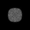
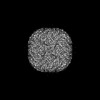
- PDB-7rvb: High resolution map of molecular chaperone Artemin -
+
Open data
ID or keywords:
Loading...
-
Basic information
Entry
Database: PDB / ID: 7rvb
Title
High resolution map of molecular chaperone Artemin
Components
Ferritin
Keywords
CHAPERONE / Molecular chaperone Artemin is a homolog of apoferritin
Function / homology
Function and homology information
ferroxidase / ferroxidase activity / ferric iron binding / iron ion transport / ferrous iron binding / intracellular iron ion homeostasis / cytoplasm Similarity search - Function
Journal: Front Mol Biosci / Year: 2022 Title: Cryo-EM structure of the diapause chaperone artemin. Authors: Amar D Parvate / Samantha M Powell / Jory T Brookreson / Trevor H Moser / Irina V Novikova / Mowei Zhou / James E Evans / Abstract: The protein artemin acts as both an RNA and protein chaperone and constitutes over 10% of all protein in cysts during diapause. However, its mechanistic details remain elusive since no high- ...The protein artemin acts as both an RNA and protein chaperone and constitutes over 10% of all protein in cysts during diapause. However, its mechanistic details remain elusive since no high-resolution structure of artemin exists. Here we report the full-length structure of artemin at 2.04 Å resolution. The cryo-EM map contains density for an intramolecular disulfide bond between Cys22-Cys61 and resolves the entire C-terminus extending into the core of the assembled protein cage but in a different configuration than previously hypothesized with molecular modeling. We also provide data supporting the role of C-terminal helix F towards stabilizing the dimer form that is believed to be important for its chaperoning activity. We were able to destabilize this effect by placing a tag at the C-terminus to fully pack the internal cavity and cause limited steric hindrance.
Average exposure time: 1.5 sec. / Electron dose: 50 e/Å2 / Film or detector model: GATAN K3 BIOQUANTUM (6k x 4k) / Num. of grids imaged: 1 / Num. of real images: 2595
EM imaging optics
Energyfilter slit width: 20 eV
-
Processing
EM software
ID
Name
Category
1
cryoSPARC
particleselection
2
EPU
imageacquisition
7
PHENIX
modelfitting
9
PHENIX
modelrefinement
13
cryoSPARC
3Dreconstruction
CTF correction
Type: PHASE FLIPPING AND AMPLITUDE CORRECTION
Particle selection
Num. of particles selected: 167408
Symmetry
Point symmetry: O (octahedral)
3D reconstruction
Resolution: 2.04 Å / Resolution method: FSC 0.143 CUT-OFF / Num. of particles: 167408 / Symmetry type: POINT
Atomic model building
Protocol: AB INITIO MODEL
+
About Yorodumi
-
News
-
Feb 9, 2022. New format data for meta-information of EMDB entries
New format data for meta-information of EMDB entries
Version 3 of the EMDB header file is now the official format.
The previous official version 1.9 will be removed from the archive.
In the structure databanks used in Yorodumi, some data are registered as the other names, "COVID-19 virus" and "2019-nCoV". Here are the details of the virus and the list of structure data.
Jan 31, 2019. EMDB accession codes are about to change! (news from PDBe EMDB page)
EMDB accession codes are about to change! (news from PDBe EMDB page)
The allocation of 4 digits for EMDB accession codes will soon come to an end. Whilst these codes will remain in use, new EMDB accession codes will include an additional digit and will expand incrementally as the available range of codes is exhausted. The current 4-digit format prefixed with “EMD-” (i.e. EMD-XXXX) will advance to a 5-digit format (i.e. EMD-XXXXX), and so on. It is currently estimated that the 4-digit codes will be depleted around Spring 2019, at which point the 5-digit format will come into force.
The EM Navigator/Yorodumi systems omit the EMD- prefix.
Related info.:Q: What is EMD? / ID/Accession-code notation in Yorodumi/EM Navigator
Yorodumi is a browser for structure data from EMDB, PDB, SASBDB, etc.
This page is also the successor to EM Navigator detail page, and also detail information page/front-end page for Omokage search.
The word "yorodu" (or yorozu) is an old Japanese word meaning "ten thousand". "mi" (miru) is to see.
Related info.:EMDB / PDB / SASBDB / Comparison of 3 databanks / Yorodumi Search / Aug 31, 2016. New EM Navigator & Yorodumi / Yorodumi Papers / Jmol/JSmol / Function and homology information / Changes in new EM Navigator and Yorodumi
 Movie
Movie Controller


 Open data
 Basic information
 Components
 Keywords
 Function and homology information
 Artemia franciscana (arthropod)
 Authors
 United States, 1items
  Citation
 Structure visualization
 Downloads & links
 Other downloads
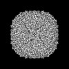
 PDBj
PDBj


 Assembly
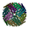

 Sample preparation
 Electron microscopy imaging
 FIELD EMISSION GUN / Accelerating voltage: 300 kV / Illumination mode: FLOOD BEAM
 Processing