Movie
Movie Controller


+ Open data

- Basic information
| Entry | Database: PDB / ID: 7rtj | ||||||
|---|---|---|---|---|---|---|---|
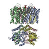
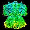
| Title | SthK Y26F Activated State | ||||||
 Components Components | SthK | ||||||
 Keywords Keywords | TRANSPORT PROTEIN / Cyclic nucleotide-gated ion channel | ||||||
| Function / homology | ADENOSINE-3',5'-CYCLIC-MONOPHOSPHATE / Chem-PGW Function and homology information Function and homology information | ||||||
| Biological species |   Spirochaeta thermophila (bacteria) Spirochaeta thermophila (bacteria) | ||||||
| Method | ELECTRON MICROSCOPY / single particle reconstruction / cryo EM / Resolution: 3.8 Å | ||||||
 Authors Authors | Gao, X. / Nimigean, C. | ||||||
| Funding support |  United States, 1items United States, 1items
| ||||||
 Citation Citation | Journal: Nat Commun / Year: 2022 Title: Gating intermediates reveal inhibitory role of the voltage sensor in a cyclic nucleotide-modulated ion channel. Authors: Xiaolong Gao / Philipp A M Schmidpeter / Vladimir Berka / Ryan J Durham / Chen Fan / Vasanthi Jayaraman / Crina M Nimigean /  Abstract: Understanding how ion channels gate is important for elucidating their physiological roles and targeting them in pathophysiological states. Here, we used SthK, a cyclic nucleotide-modulated channel ...Understanding how ion channels gate is important for elucidating their physiological roles and targeting them in pathophysiological states. Here, we used SthK, a cyclic nucleotide-modulated channel from Spirochaeta thermophila, to define a ligand-gating trajectory that includes multiple on-pathway intermediates. cAMP is a poor partial agonist for SthK and depolarization increases SthK activity. Tuning the energy landscape by gain-of-function mutations in the voltage sensor domain (VSD) allowed us to capture multiple intermediates along the ligand-activation pathway, highlighting the allosteric linkage between VSD, cyclic nucleotide-binding (CNBD) and pore domains. Small, lateral displacements of the VSD S4 segment were necessary to open the intracellular gate, pointing to an inhibitory VSD at rest. We propose that in wild-type SthK, depolarization leads to such VSD displacements resulting in release of inhibition. In summary, we report conformational transitions along the activation pathway that reveal allosteric couplings between key sites integrating to open the intracellular gate. | ||||||
| History |
|
- Structure visualization
| Structure viewer | Molecule: MolmilJmol/JSmol |
|---|
- Downloads & links
-Download
| PDBx/mmCIF format | 7rtj.cif.gz | 355.1 KB | Display | PDBx/mmCIF format |
|---|---|---|---|---|
| PDB format | pdb7rtj.ent.gz | 282 KB | Display | PDB format |
| PDBx/mmJSON format | 7rtj.json.gz | Tree view | PDBx/mmJSON format | |
| Others |  Other downloads Other downloads |
-Validation report
| Summary document | 7rtj_validation.pdf.gz | 1.5 MB | Display | wwPDB validaton report |
|---|---|---|---|---|
| Full document | 7rtj_full_validation.pdf.gz | 1.6 MB | Display | |
| Data in XML | 7rtj_validation.xml.gz | 57.1 KB | Display | |
| Data in CIF | 7rtj_validation.cif.gz | 80.8 KB | Display | |
| Arichive directory | https://data.pdbj.org/pub/pdb/validation_reports/rt/7rtjftp://data.pdbj.org/pub/pdb/validation_reports/rt/7rtj | HTTPS FTP |
-Related structure data
| Related structure data | 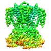 24682MC  7rshC  7rtfC  7ru0C  7ryrC  7rysC C: citing same article ( M: map data used to model this data |
|---|---|
| Similar structure data |


-Links
 PDBj
PDBj


- Assembly
| Deposited unit | 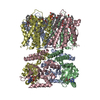
|
|---|---|
| 1 |
|
-Components
| #1: Protein | Mass: 51102.574 Da / Num. of mol.: 4 / Mutation: Y26F Source method: isolated from a genetically manipulated source Source: (gene. exp.) Spirochaeta thermophila (bacteria) / Production host: #2: Chemical | ChemComp-CMP /   Mass: 329.206 Da / Num. of mol.: 4 / Source method: obtained synthetically / Formula: C10H12N5O6P / Feature type: SUBJECT OF INVESTIGATION Mass: 329.206 Da / Num. of mol.: 4 / Source method: obtained synthetically / Formula: C10H12N5O6P / Feature type: SUBJECT OF INVESTIGATION#3: Chemical | ChemComp-PGW / (   Mass: 749.007 Da / Num. of mol.: 32 / Source method: obtained synthetically / Formula: C40H77O10P / Comment: phospholipid*YM Mass: 749.007 Da / Num. of mol.: 32 / Source method: obtained synthetically / Formula: C40H77O10P / Comment: phospholipid*YMHas ligand of interest | Y | |
|---|
-Experimental details
-Experiment
| Experiment | Method: ELECTRON MICROSCOPY |
|---|---|
| EM experiment | Aggregation state: PARTICLE / 3D reconstruction method: single particle reconstruction |
- Sample preparation
| Component | Name: SthK / Type: COMPLEX / Details: Y26F mutant of wild type SthK / Entity ID: #1 / Source: RECOMBINANT | |||||||||||||||
|---|---|---|---|---|---|---|---|---|---|---|---|---|---|---|---|---|
| Molecular weight | Experimental value: NO | |||||||||||||||
| Source (natural) | Organism: Spirochaeta thermophila (bacteria) | |||||||||||||||
| Source (recombinant) | Organism: | |||||||||||||||
| Buffer solution | pH: 7 | |||||||||||||||
| Buffer component |
| |||||||||||||||
| Specimen | Conc.: 7.5 mg/ml / Embedding applied: NO / Shadowing applied: NO / Staining applied: NO / Vitrification applied: YES / Details: Protein in nanodisc of DOPC/POPG/Cardiolipin | |||||||||||||||
| Specimen support | Grid material: GOLD / Grid mesh size: 300 divisions/in. / Grid type: Quantifoil R1.2/1.3 | |||||||||||||||
| Vitrification | Instrument: FEI VITROBOT MARK IV / Cryogen name: ETHANE / Humidity: 100 % / Chamber temperature: 295 K |
- Electron microscopy imaging
| Experimental equipment |  Model: Titan Krios / Image courtesy: FEI Company |
|---|---|
| Microscopy | Model: FEI TITAN KRIOS |
| Electron gun | Electron source:  FIELD EMISSION GUN / Accelerating voltage: 300 kV / Illumination mode: FLOOD BEAM FIELD EMISSION GUN / Accelerating voltage: 300 kV / Illumination mode: FLOOD BEAM |
| Electron lens | Mode: BRIGHT FIELD / Nominal magnification: 130000 X / Nominal defocus max: 3000 nm / Nominal defocus min: 1000 nm / Cs: 2.7 mm |
| Specimen holder | Cryogen: NITROGEN / Specimen holder model: FEI TITAN KRIOS AUTOGRID HOLDER |
| Image recording | Electron dose: 1.365 e/Å2 / Detector mode: SUPER-RESOLUTION / Film or detector model: GATAN K2 SUMMIT (4k x 4k) |
| Image scans | Movie frames/image: 40 |
- Processing
| Software | Name: PHENIX / Version: 1.20.1_4487: / Classification: refinement | ||||||||||||||||||||||||||||||||||||
|---|---|---|---|---|---|---|---|---|---|---|---|---|---|---|---|---|---|---|---|---|---|---|---|---|---|---|---|---|---|---|---|---|---|---|---|---|---|
| EM software |
| ||||||||||||||||||||||||||||||||||||
| CTF correction | Type: PHASE FLIPPING AND AMPLITUDE CORRECTION | ||||||||||||||||||||||||||||||||||||
| Particle selection | Num. of particles selected: 1324724 | ||||||||||||||||||||||||||||||||||||
| Symmetry | Point symmetry: C4 (4 fold cyclic) | ||||||||||||||||||||||||||||||||||||
| 3D reconstruction | Resolution: 3.8 Å / Resolution method: FSC 0.143 CUT-OFF / Num. of particles: 109647 / Symmetry type: POINT | ||||||||||||||||||||||||||||||||||||
| Atomic model building | Space: REAL | ||||||||||||||||||||||||||||||||||||
| Atomic model building | PDB-ID: 6CJU Accession code: 6CJU / Source name: PDB / Type: experimental model | ||||||||||||||||||||||||||||||||||||
| Refine LS restraints |
|