 Movie
Movie Controller


+ Open data

- Basic information
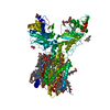
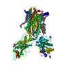
| Entry | Database: PDB / ID: 7rpi | ||||||||||||||||||
|---|---|---|---|---|---|---|---|---|---|---|---|---|---|---|---|---|---|---|---|
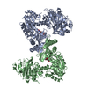
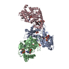
| Title | Cryo-EM structure of murine Dispatched 'T' conformation | ||||||||||||||||||
 Components Components | Protein dispatched homolog 1 | ||||||||||||||||||
 Keywords Keywords | MEMBRANE PROTEIN / RND transporter / Hedgehog binding / Sterol sensing domain / sodium binding | ||||||||||||||||||
| Function / homology |  Function and homology information Function and homology informationpatched ligand maturation / diaphragm development / embryonic pattern specification / dorsal/ventral pattern formation / peptide transport / determination of left/right symmetry / molecular carrier activity / smoothened signaling pathway / membrane Similarity search - Function | ||||||||||||||||||
| Biological species |  | ||||||||||||||||||
| Method | ELECTRON MICROSCOPY / single particle reconstruction / cryo EM / Resolution: 2.5 Å | ||||||||||||||||||
 Authors Authors | Asarnow, D. / Wang, Q. / Ding, K. / Cheng, Y. / Beachy, P.A. | ||||||||||||||||||
| Funding support |  United States, 5items United States, 5items
| ||||||||||||||||||
 Citation Citation | Journal: Nature / Year: 2021 Title: Dispatched uses Na flux to power release of lipid-modified Hedgehog. Authors: Qianqian Wang / Daniel E Asarnow / Ke Ding / Randall K Mann / Jason Hatakeyama / Yunxiao Zhang / Yong Ma / Yifan Cheng / Philip A Beachy /  Abstract: The Dispatched protein, which is related to the NPC1 and PTCH1 cholesterol transporters and to H-driven transporters of the RND family, enables tissue-patterning activity of the lipid-modified ...The Dispatched protein, which is related to the NPC1 and PTCH1 cholesterol transporters and to H-driven transporters of the RND family, enables tissue-patterning activity of the lipid-modified Hedgehog protein by releasing it from tightly -localized sites of embryonic expression. Here we determine a cryo-electron microscopy structure of the mouse protein Dispatched homologue 1 (DISP1), revealing three Na ions coordinated within a channel that traverses its transmembrane domain. We find that the rate of Hedgehog export is dependent on the Na gradient across the plasma membrane. The transmembrane channel and Na binding are disrupted in DISP1-NNN, a variant with asparagine substitutions for three intramembrane aspartate residues that each coordinate and neutralize the charge of one of the three Na ions. DISP1-NNN and variants that disrupt single Na sites retain binding to, but are impaired in export of the lipid-modified Hedgehog protein to the SCUBE2 acceptor. Interaction of the amino-terminal signalling domain of the Sonic hedgehog protein (ShhN) with DISP1 occurs via an extensive buried surface area and contacts with an extended furin-cleaved DISP1 arm. Variability analysis reveals that ShhN binding is restricted to one extreme of a continuous series of DISP1 conformations. The bound and unbound DISP1 conformations display distinct Na-site occupancies, which suggests a mechanism by which transmembrane Na flux may power extraction of the lipid-linked Hedgehog signal from the membrane. Na-coordinating residues in DISP1 are conserved in PTCH1 and other metazoan RND family members, suggesting that Na flux powers their conformationally driven activities. | ||||||||||||||||||
| History |
|
- Structure visualization
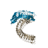
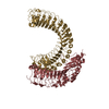
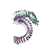
| Movie |
Movie viewer |
|---|---|
| Structure viewer | Molecule: MolmilJmol/JSmol |
- Downloads & links
-Download
| PDBx/mmCIF format | 7rpi.cif.gz | 211.8 KB | Display | PDBx/mmCIF format |
|---|---|---|---|---|
| PDB format | pdb7rpi.ent.gz | 163.5 KB | Display | PDB format |
| PDBx/mmJSON format | 7rpi.json.gz | Tree view | PDBx/mmJSON format | |
| Others |  Other downloads Other downloads |
-Validation report
| Arichive directory | https://data.pdbj.org/pub/pdb/validation_reports/rp/7rpiftp://data.pdbj.org/pub/pdb/validation_reports/rp/7rpi | HTTPS FTP |
|---|
-Related structure data
| Related structure data |  24615MC  7rphC 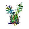 7rpjC  7rpkC M: map data used to model this data C: citing same article ( |
|---|---|
| Similar structure data |


-Links
 PDBj
PDBj
- Assembly
| Deposited unit | 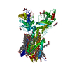
|
|---|---|
| 1 |
|
-Components
-Protein / Sugars , 2 types, 6 molecules A
| #1: Protein | Mass: 152071.141 Da / Num. of mol.: 1 / Fragment: UNP residues 172-1521 Source method: isolated from a genetically manipulated source Source: (gene. exp.)  Homo sapiens (human) / References: UniProt: Q3TDN0 Homo sapiens (human) / References: UniProt: Q3TDN0 |
|---|---|
| #4: Sugar | ChemComp-NAG /  Type: D-saccharide, beta linking / Mass: 221.208 Da / Num. of mol.: 5 / Source method: obtained synthetically / Formula: C8H15NO6 Type: D-saccharide, beta linking / Mass: 221.208 Da / Num. of mol.: 5 / Source method: obtained synthetically / Formula: C8H15NO6 |
-Non-polymers , 4 types, 73 molecules 


| #2: Chemical | ChemComp-Y01 /  Mass: 486.726 Da / Num. of mol.: 26 / Source method: obtained synthetically / Formula: C31H50O4 / Feature type: SUBJECT OF INVESTIGATION Mass: 486.726 Da / Num. of mol.: 26 / Source method: obtained synthetically / Formula: C31H50O4 / Feature type: SUBJECT OF INVESTIGATION#3: Chemical |  Mass: 1005.188 Da / Num. of mol.: 2 / Source method: obtained synthetically / Formula: C47H88O22 / Feature type: SUBJECT OF INVESTIGATION Mass: 1005.188 Da / Num. of mol.: 2 / Source method: obtained synthetically / Formula: C47H88O22 / Feature type: SUBJECT OF INVESTIGATION#5: Chemical |  Mass: 22.990 Da / Num. of mol.: 2 / Source method: obtained synthetically / Formula: Na / Feature type: SUBJECT OF INVESTIGATION Mass: 22.990 Da / Num. of mol.: 2 / Source method: obtained synthetically / Formula: Na / Feature type: SUBJECT OF INVESTIGATION#6: Water | ChemComp-HOH / | Mass: 18.015 Da / Num. of mol.: 43 / Source method: isolated from a natural source / Formula: H2O |
|---|
-Details
| Has ligand of interest | Y |
|---|---|
| Has protein modification | Y |
-Experimental details
-Experiment
| Experiment | Method: ELECTRON MICROSCOPY |
|---|---|
| EM experiment | Aggregation state: PARTICLE / 3D reconstruction method: single particle reconstruction |
- Sample preparation
| Component | Name: Dispatched protein 'T' conformation / Type: COMPLEX / Entity ID: #1 / Source: RECOMBINANT | |||||||||||||||||||||||||||||||||||
|---|---|---|---|---|---|---|---|---|---|---|---|---|---|---|---|---|---|---|---|---|---|---|---|---|---|---|---|---|---|---|---|---|---|---|---|---|
| Molecular weight | Value: 0.15191493 MDa / Experimental value: NO | |||||||||||||||||||||||||||||||||||
| Source (natural) | Organism: | |||||||||||||||||||||||||||||||||||
| Source (recombinant) | Organism: Homo sapiens (human) / Strain: HEK293F | |||||||||||||||||||||||||||||||||||
| Buffer solution | pH: 7.5 | |||||||||||||||||||||||||||||||||||
| Buffer component |
| |||||||||||||||||||||||||||||||||||
| Specimen | Conc.: 1.56 mg/ml / Embedding applied: NO / Shadowing applied: NO / Staining applied: NO / Vitrification applied: YES | |||||||||||||||||||||||||||||||||||
| Vitrification | Instrument: FEI VITROBOT MARK IV / Cryogen name: ETHANE / Humidity: 100 % / Chamber temperature: 281 K Details: Wait time 20 seconds, blot time 4 seconds, blotting force 0 |
- Electron microscopy imaging
| Experimental equipment |  Model: Titan Krios / Image courtesy: FEI Company |
|---|---|
| Microscopy | Model: FEI TITAN KRIOS Details: Automated data collection in SerialEM using 3x3 image shift pattern (one shot per hole), using beam tilt compensation. |
| Electron gun | Electron source:  FIELD EMISSION GUN / Accelerating voltage: 300 kV / Illumination mode: FLOOD BEAM FIELD EMISSION GUN / Accelerating voltage: 300 kV / Illumination mode: FLOOD BEAM |
| Electron lens | Mode: BRIGHT FIELD / Nominal magnification: 105000 X / Calibrated magnification: 59880 X / Nominal defocus max: 1500 nm / Nominal defocus min: 500 nm / Cs: 2.7 mm / C2 aperture diameter: 100 µm / Alignment procedure: COMA FREE |
| Specimen holder | Cryogen: NITROGEN / Specimen holder model: FEI TITAN KRIOS AUTOGRID HOLDER |
| Image recording | Electron dose: 66.7 e/Å2 / Film or detector model: GATAN K3 BIOQUANTUM (6k x 4k) / Num. of grids imaged: 1 / Num. of real images: 4487 |
| EM imaging optics | Energyfilter name: GIF Bioquantum / Energyfilter slit width: 20 eV |
- Processing
| EM software |
| |||||||||||||||||||||||||||||||||||||||
|---|---|---|---|---|---|---|---|---|---|---|---|---|---|---|---|---|---|---|---|---|---|---|---|---|---|---|---|---|---|---|---|---|---|---|---|---|---|---|---|---|
| CTF correction | Type: PHASE FLIPPING AND AMPLITUDE CORRECTION | |||||||||||||||||||||||||||||||||||||||
| Particle selection | Num. of particles selected: 3717941 | |||||||||||||||||||||||||||||||||||||||
| Symmetry | Point symmetry: C1 (asymmetric) | |||||||||||||||||||||||||||||||||||||||
| 3D reconstruction | Resolution: 2.5 Å / Resolution method: FSC 0.143 CUT-OFF / Num. of particles: 154908 / Algorithm: FOURIER SPACE / Num. of class averages: 1 / Symmetry type: POINT | |||||||||||||||||||||||||||||||||||||||
| Atomic model building | B value: 72.1 / Protocol: FLEXIBLE FIT / Space: REAL |
