+ データを開く
- 基本情報
基本情報
| 登録情報 | データベース: PDB / ID: 7rm5 | |||||||||||||||
|---|---|---|---|---|---|---|---|---|---|---|---|---|---|---|---|---|
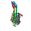
| タイトル | MicroED structure of the human adenosine receptor at 2.8A | |||||||||||||||
 要素 要素 | Adenosine receptor A2a/Soluble cytochrome b562 chimera | |||||||||||||||
 キーワード キーワード | MEMBRANE PROTEIN | |||||||||||||||
| 機能・相同性 |  機能・相同性情報 機能・相同性情報positive regulation of acetylcholine secretion, neurotransmission / positive regulation of circadian sleep/wake cycle, sleep / regulation of norepinephrine secretion / negative regulation of alpha-beta T cell activation / Adenosine P1 receptors / G protein-coupled adenosine receptor activity / G protein-coupled adenosine receptor signaling pathway / response to purine-containing compound / sensory perception / NGF-independant TRKA activation ...positive regulation of acetylcholine secretion, neurotransmission / positive regulation of circadian sleep/wake cycle, sleep / regulation of norepinephrine secretion / negative regulation of alpha-beta T cell activation / Adenosine P1 receptors / G protein-coupled adenosine receptor activity / G protein-coupled adenosine receptor signaling pathway / response to purine-containing compound / sensory perception / NGF-independant TRKA activation / Surfactant metabolism / positive regulation of urine volume / positive regulation of glutamate secretion / synaptic transmission, dopaminergic / inhibitory postsynaptic potential / negative regulation of vascular permeability / : / type 5 metabotropic glutamate receptor binding / synaptic transmission, cholinergic / blood circulation / response to caffeine / intermediate filament / eating behavior / alpha-actinin binding / presynaptic active zone / regulation of calcium ion transport / membrane depolarization / asymmetric synapse / axolemma / cellular defense response / prepulse inhibition / phagocytosis / presynaptic modulation of chemical synaptic transmission / response to amphetamine / positive regulation of synaptic transmission, glutamatergic / neuron projection morphogenesis / excitatory postsynaptic potential / regulation of mitochondrial membrane potential / apoptotic signaling pathway / positive regulation of long-term synaptic potentiation / synaptic transmission, glutamatergic / central nervous system development / positive regulation of synaptic transmission, GABAergic / positive regulation of protein secretion / locomotory behavior / astrocyte activation / positive regulation of apoptotic signaling pathway / electron transport chain / adenylate cyclase-modulating G protein-coupled receptor signaling pathway / adenylate cyclase-activating G protein-coupled receptor signaling pathway / negative regulation of inflammatory response / vasodilation / blood coagulation / cell-cell signaling / presynaptic membrane / G alpha (s) signalling events / postsynaptic membrane / negative regulation of neuron apoptotic process / periplasmic space / electron transfer activity / calmodulin binding / inflammatory response / iron ion binding / response to xenobiotic stimulus / negative regulation of cell population proliferation / neuronal cell body / lipid binding / glutamatergic synapse / dendrite / heme binding / protein-containing complex binding / regulation of DNA-templated transcription / apoptotic process / enzyme binding / identical protein binding / membrane / plasma membrane 類似検索 - 分子機能 | |||||||||||||||
| 生物種 |  Homo sapiens (ヒト) Homo sapiens (ヒト) | |||||||||||||||
| 手法 | 電子線結晶学 /  分子置換 / クライオ電子顕微鏡法 / 解像度: 2.79 Å 分子置換 / クライオ電子顕微鏡法 / 解像度: 2.79 Å | |||||||||||||||
 データ登録者 データ登録者 | Martynowycz, M.W. / Shiriaeva, A. / Ge, X. / Hattne, J. / Nannenga, B.L. / Cherezov, V. / Gonen, T. | |||||||||||||||
| 資金援助 |  米国, 4件 米国, 4件
| |||||||||||||||
 引用 引用 |  ジャーナル: Proc Natl Acad Sci U S A / 年: 2021 ジャーナル: Proc Natl Acad Sci U S A / 年: 2021タイトル: MicroED structure of the human adenosine receptor determined from a single nanocrystal in LCP. 著者: Michael W Martynowycz / Anna Shiriaeva / Xuanrui Ge / Johan Hattne / Brent L Nannenga / Vadim Cherezov / Tamir Gonen /  要旨: G protein-coupled receptors (GPCRs), or seven-transmembrane receptors, are a superfamily of membrane proteins that are critically important to physiological processes in the human body. Determining ...G protein-coupled receptors (GPCRs), or seven-transmembrane receptors, are a superfamily of membrane proteins that are critically important to physiological processes in the human body. Determining high-resolution structures of GPCRs without bound cognate signaling partners, such as a G protein, requires crystallization in lipidic cubic phase (LCP). GPCR crystals grown in LCP are often too small for traditional X-ray crystallography. These microcrystals are ideal for investigation by microcrystal electron diffraction (MicroED), but the gel-like nature of LCP makes traditional approaches to MicroED sample preparation insurmountable. Here, we show that the structure of a human A adenosine receptor can be determined by MicroED after converting the LCP into the sponge phase followed by focused ion-beam milling. We determined the structure of the A adenosine receptor to 2.8-Å resolution and resolved an antagonist in its orthosteric ligand-binding site, as well as four cholesterol molecules bound around the receptor. This study lays the groundwork for future structural studies of lipid-embedded membrane proteins by MicroED using single microcrystals that would be impossible with other crystallographic methods. | |||||||||||||||
| 履歴 |
|
- 構造の表示
構造の表示
| ムービー |
 ムービービューア ムービービューア |
|---|---|
| 構造ビューア | 分子:  Molmil Molmil Jmol/JSmol Jmol/JSmol |
- ダウンロードとリンク
- ダウンロード
| PDBx/mmCIF形式 |  7rm5.cif.gz 7rm5.cif.gz | 98.1 KB | 表示 |  PDBx/mmCIF形式 PDBx/mmCIF形式 |
|---|---|---|---|---|
| PDB形式 |  pdb7rm5.ent.gz pdb7rm5.ent.gz | 68.5 KB | 表示 |  PDB形式 PDB形式 |
| PDBx/mmJSON形式 |  7rm5.json.gz 7rm5.json.gz | ツリー表示 |  PDBx/mmJSON形式 PDBx/mmJSON形式 | |
| その他 |  その他のダウンロード その他のダウンロード |
-検証レポート
| 文書・要旨 |  7rm5_validation.pdf.gz 7rm5_validation.pdf.gz | 1.1 MB | 表示 |  wwPDB検証レポート wwPDB検証レポート |
|---|---|---|---|---|
| 文書・詳細版 |  7rm5_full_validation.pdf.gz 7rm5_full_validation.pdf.gz | 1.1 MB | 表示 | |
| XML形式データ |  7rm5_validation.xml.gz 7rm5_validation.xml.gz | 17.1 KB | 表示 | |
| CIF形式データ |  7rm5_validation.cif.gz 7rm5_validation.cif.gz | 23.9 KB | 表示 | |
| アーカイブディレクトリ |  https://data.pdbj.org/pub/pdb/validation_reports/rm/7rm5 https://data.pdbj.org/pub/pdb/validation_reports/rm/7rm5 ftp://data.pdbj.org/pub/pdb/validation_reports/rm/7rm5 ftp://data.pdbj.org/pub/pdb/validation_reports/rm/7rm5 | HTTPS FTP |
-関連構造データ
| 関連構造データ |  24551MC  4eiyS |
|---|---|
| 類似構造データ | 類似検索 - 機能・相同性  F&H 検索 F&H 検索 |
- リンク
リンク
- 集合体
集合体
| 登録構造単位 | 
| ||||||||||||
|---|---|---|---|---|---|---|---|---|---|---|---|---|---|
| 1 | 
| ||||||||||||
| 単位格子 |
|
- 要素
要素
| #1: タンパク質 | 分子量: 49974.281 Da / 分子数: 1 / 由来タイプ: 組換発現 由来: (組換発現)  Homo sapiens (ヒト), (組換発現) Homo sapiens (ヒト), (組換発現)  遺伝子: ADORA2A, ADORA2, cybC 発現宿主:  参照: UniProt: P29274, UniProt: P0ABE7 | ||||||
|---|---|---|---|---|---|---|---|
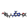
| #2: 化合物 | ChemComp-ZMA / | ||||||
| #3: 化合物 | ChemComp-CLR / #4: 化合物 | ChemComp-NA / | #5: 水 | ChemComp-HOH / | 研究の焦点であるリガンドがあるか | N | |
-実験情報
-実験
| 実験 | 手法: 電子線結晶学 |
|---|---|
| EM実験 | 試料の集合状態: 3D ARRAY / 3次元再構成法: 電子線結晶学 |
- 試料調製
試料調製
| 構成要素 | 名称: Adenosine receptor A2a/Soluble cytochrome b562 chimera タイプ: COMPLEX / Entity ID: #1 / 由来: RECOMBINANT | ||||||||||||
|---|---|---|---|---|---|---|---|---|---|---|---|---|---|
| 分子量 | 値: 0.05872 MDa / 実験値: NO | ||||||||||||
| 由来(天然) |
| ||||||||||||
| 由来(組換発現) |
| ||||||||||||
| 緩衝液 | pH: 5 詳細: 25-28% (v/v) PEG 400, 0.04-0.06M sodium thiocyanate, 2% (v/v) 2,5-hexanediol, 100mM sodium citrate, pH 5.0, Lipid Cubic Phase (LCP), temperature 293K | ||||||||||||
| 試料 | 濃度: 10 mg/ml / 包埋: NO / シャドウイング: NO / 染色: NO / 凍結: YES | ||||||||||||
| 試料支持 | グリッドの材料: COPPER / グリッドのサイズ: 200 divisions/in. / グリッドのタイプ: Quantifoil R2/2 | ||||||||||||
| 急速凍結 | 装置: LEICA EM GP / 凍結剤: ETHANE / 湿度: 95 % / 凍結前の試料温度: 298 K |
-データ収集
| 実験機器 |  モデル: Titan Krios / 画像提供: FEI Company |
|---|---|
| 顕微鏡 | モデル: FEI TITAN KRIOS |
| 電子銃 | 電子線源:  FIELD EMISSION GUN / 加速電圧: 300 kV / 照射モード: FLOOD BEAM FIELD EMISSION GUN / 加速電圧: 300 kV / 照射モード: FLOOD BEAM |
| 電子レンズ | モード: DIFFRACTION / 最大 デフォーカス(公称値): 0 nm / 最小 デフォーカス(公称値): 0 nm / Cs: 2.7 mm / C2レンズ絞り径: 50 µm / アライメント法: BASIC |
| 試料ホルダ | 凍結剤: NITROGEN 試料ホルダーモデル: FEI TITAN KRIOS AUTOGRID HOLDER 最高温度: 83 K / 最低温度: 77 K |
| 撮影 | 平均露光時間: 3 sec. / 電子線照射量: 0.01 e/Å2 / フィルム・検出器のモデル: FEI CETA (4k x 4k) / Num. of diffraction images: 134 / 撮影したグリッド数: 1 |
| 画像スキャン | サンプリングサイズ: 28 µm / 横: 2048 / 縦: 2048 |
| EM回折 | カメラ長: 1900 mm / Tilt angle list: -40, +40, 0.6 |
| EM回折 シェル | 解像度: 37.91→2.79 Å / フーリエ空間範囲: 77 % / 多重度: 3.7 / 構造因子数: 10071 / 位相残差: 30 ° |
| EM回折 統計 | フーリエ空間範囲: 77 % / 再高解像度: 2.79 Å / 測定した強度の数: 37130 / 構造因子数: 10071 / 位相誤差: 30 ° / 位相誤差の除外基準: none / Rmerge: 0.299 |
| 回折 | Serial crystal experiment: N |
| 検出器 | 検出器: CMOS / 日付: 2020年9月11日 |
- 解析
解析
| ソフトウェア |
| |||||||||||||||||||||
|---|---|---|---|---|---|---|---|---|---|---|---|---|---|---|---|---|---|---|---|---|---|---|
| EMソフトウェア |
| |||||||||||||||||||||
| 画像処理 | 詳細: CetaD binned by 2 | |||||||||||||||||||||
| EM 3D crystal entity | ∠α: 90 ° / ∠β: 90 ° / ∠γ: 90 ° / A: 40 Å / B: 180.5 Å / C: 139.7 Å / 空間群名: C2221 / 空間群番号: 20 | |||||||||||||||||||||
| CTF補正 | タイプ: NONE | |||||||||||||||||||||
| 3次元再構成 | 解像度: 2.79 Å / 解像度の算出法: DIFFRACTION PATTERN/LAYERLINES / アルゴリズム: FOURIER SPACE / 対称性のタイプ: 3D CRYSTAL | |||||||||||||||||||||
| 原子モデル構築 | B value: 43 / プロトコル: RIGID BODY FIT / 空間: RECIPROCAL / Target criteria: Maximum likelihood | |||||||||||||||||||||
| 原子モデル構築 | PDB-ID: 4EIY PDB chain-ID: A / Accession code: 4EIY / Source name: PDB / タイプ: experimental model | |||||||||||||||||||||
| 精密化 | 構造決定の手法:  分子置換 分子置換開始モデル: 4EIY 解像度: 2.79→37.91 Å / SU B: 0.48 / 交差検証法: FREE R-VALUE / σ(F): 1.3 / 位相誤差: 0.3063
| |||||||||||||||||||||
| 溶媒の処理 | 減衰半径: 0.9 Å / VDWプローブ半径: 1.11 Å | |||||||||||||||||||||
| 原子変位パラメータ | Biso mean: 43 Å2 | |||||||||||||||||||||
| LS精密化 シェル | 解像度: 2.79→3.07 Å
|
 ムービー
ムービー コントローラー


 PDBj
PDBj


