 Movie
Movie Controller


+ Open data

- Basic information
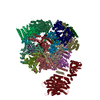
| Entry | Database: PDB / ID: 7qve | ||||||
|---|---|---|---|---|---|---|---|
| Title | Spinach 20S proteasome | ||||||
 Components Components | (Proteasome subunit ...) x 14 | ||||||
 Keywords Keywords | PLANT PROTEIN / PROTEASOME / UPS / PLANT / SPINACH | ||||||
| Function / homology |  Function and homology information Function and homology informationproteasome core complex / proteasome endopeptidase complex / proteasome core complex, beta-subunit complex / threonine-type endopeptidase activity / proteasome core complex, alpha-subunit complex / proteasomal protein catabolic process / peptidase activity / endopeptidase activity / ubiquitin-dependent protein catabolic process / proteasome-mediated ubiquitin-dependent protein catabolic process ...proteasome core complex / proteasome endopeptidase complex / proteasome core complex, beta-subunit complex / threonine-type endopeptidase activity / proteasome core complex, alpha-subunit complex / proteasomal protein catabolic process / peptidase activity / endopeptidase activity / ubiquitin-dependent protein catabolic process / proteasome-mediated ubiquitin-dependent protein catabolic process / nucleus / cytoplasm / cytosol Similarity search - Function | ||||||
| Biological species |  Spinacia oleracea (spinach) Spinacia oleracea (spinach) | ||||||
| Method | ELECTRON MICROSCOPY / single particle reconstruction / cryo EM / Resolution: 3.3 Å | ||||||
 Authors Authors | Kandolf, S. / Grishkovskaya, I. / Meinhart, A. / Haselbach, D. | ||||||
| Funding support | 1items
| ||||||
 Citation Citation | Journal: Plant Commun / Year: 2022 Title: Cryo-EM structure of the plant 26S proteasome. Authors: Susanne Kandolf / Irina Grishkovskaya / Katarina Belačić / Derek L Bolhuis / Sascha Amann / Brent Foster / Richard Imre / Karl Mechtler / Alexander Schleiffer / Hemant D Tagare / Ellen D ...Authors: Susanne Kandolf / Irina Grishkovskaya / Katarina Belačić / Derek L Bolhuis / Sascha Amann / Brent Foster / Richard Imre / Karl Mechtler / Alexander Schleiffer / Hemant D Tagare / Ellen D Zhong / Anton Meinhart / Nicholas G Brown / David Haselbach /    Abstract: Targeted proteolysis is a hallmark of life. It is especially important in long-lived cells that can be found in higher eukaryotes, like plants. This task is mainly fulfilled by the ubiquitin- ...Targeted proteolysis is a hallmark of life. It is especially important in long-lived cells that can be found in higher eukaryotes, like plants. This task is mainly fulfilled by the ubiquitin-proteasome system. Thus, proteolysis by the 26S proteasome is vital to development, immunity, and cell division. Although the yeast and animal proteasomes are well characterized, there is only limited information on the plant proteasome. We determined the first plant 26S proteasome structure from Spinacia oleracea by single-particle electron cryogenic microscopy at an overall resolution of 3.3 Å. We found an almost identical overall architecture of the spinach proteasome compared with the known structures from mammals and yeast. Nevertheless, we noticed a structural difference in the proteolytic active β1 subunit. Furthermore, we uncovered an unseen compression state by characterizing the proteasome's conformational landscape. We suspect that this new conformation of the 20S core protease, in correlation with a partial opening of the unoccupied gate, may contribute to peptide release after proteolysis. Our data provide a structural basis for the plant proteasome, which is crucial for further studies. | ||||||
| History |
|
- Structure visualization
| Structure viewer | Molecule: MolmilJmol/JSmol |
|---|
- Downloads & links
-Download
| PDBx/mmCIF format | 7qve.cif.gz | 1 MB | Display | PDBx/mmCIF format |
|---|---|---|---|---|
| PDB format | pdb7qve.ent.gz | 863.5 KB | Display | PDB format |
| PDBx/mmJSON format | 7qve.json.gz | Tree view | PDBx/mmJSON format | |
| Others |  Other downloads Other downloads |
-Validation report
| Arichive directory | https://data.pdbj.org/pub/pdb/validation_reports/qv/7qveftp://data.pdbj.org/pub/pdb/validation_reports/qv/7qve | HTTPS FTP |
|---|
-Related structure data
| Related structure data |  14175MC M: map data used to model this data C: citing same article ( |
|---|---|
| Similar structure data | |
| EM raw data | EMPIAR-10974 (Title: Cryo-EM structure of the plant 26S proteasome / Data size: 7.9 TB Data #1: Unaligned multi-frame micrographs of spinach 26S proteasome [micrographs - multiframe] Data #2: Unaligned multi-frame micrographs of spinach 26S proteasome [micrographs - multiframe] Data #3: Unaligned multi-frame micrographs of spinach 26S proteasome [micrographs - multiframe]) |
-Links
 PDBj
PDBj


- Assembly
| Deposited unit | 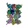
|
|---|---|
| 1 |
|
-Components
-Proteasome subunit ... , 14 types, 28 molecules iCjDkElFmGnXBhoapbqcrdsetfug
| #1: Protein | Mass: 25588.023 Da / Num. of mol.: 2 / Source method: isolated from a natural source / Source: (natural) Spinacia oleracea (spinach) / References: UniProt: A0A0K9QA79#2: Protein | Mass: 27504.117 Da / Num. of mol.: 2 / Source method: isolated from a natural source / Source: (natural) Spinacia oleracea (spinach) / References: UniProt: A0A0K9RH20#3: Protein | Mass: 26844.439 Da / Num. of mol.: 2 / Source method: isolated from a natural source / Source: (natural) Spinacia oleracea (spinach) / References: UniProt: A0A0K9S0K6#4: Protein | Mass: 26027.238 Da / Num. of mol.: 2 / Source method: isolated from a natural source / Source: (natural) Spinacia oleracea (spinach) / References: UniProt: A0A0K9RE77#5: Protein | Mass: 29932.393 Da / Num. of mol.: 2 / Source method: isolated from a natural source / Source: (natural) Spinacia oleracea (spinach) / References: UniProt: A0A0K9RGG6#6: Protein | Mass: 27322.258 Da / Num. of mol.: 2 / Source method: isolated from a natural source / Source: (natural) Spinacia oleracea (spinach) / References: UniProt: O24362#7: Protein | Mass: 27211.789 Da / Num. of mol.: 2 / Source method: isolated from a natural source / Source: (natural) Spinacia oleracea (spinach) / References: UniProt: A0A0K9R8Z8#8: Protein | Mass: 25324.434 Da / Num. of mol.: 2 / Source method: isolated from a natural source / Source: (natural) Spinacia oleracea (spinach) / References: UniProt: A0A0K9S2G1#9: Protein | Mass: 29404.385 Da / Num. of mol.: 2 / Source method: isolated from a natural source / Source: (natural) Spinacia oleracea (spinach) / References: UniProt: A0A0K9S3A8#10: Protein | Mass: 22847.109 Da / Num. of mol.: 2 / Source method: isolated from a natural source / Source: (natural) Spinacia oleracea (spinach) / References: UniProt: A0A0K9S024#11: Protein | Mass: 22950.082 Da / Num. of mol.: 2 / Source method: isolated from a natural source / Source: (natural) Spinacia oleracea (spinach) / References: UniProt: A0A0K9QSM9#12: Protein | Mass: 29652.654 Da / Num. of mol.: 2 / Source method: isolated from a natural source / Source: (natural) Spinacia oleracea (spinach)References: UniProt: O24361, proteasome endopeptidase complex #13: Protein | Mass: 24835.199 Da / Num. of mol.: 2 / Source method: isolated from a natural source / Source: (natural) Spinacia oleracea (spinach) / References: UniProt: A0A0K9RTN8#14: Protein | Mass: 27098.770 Da / Num. of mol.: 2 / Source method: isolated from a natural source / Source: (natural) Spinacia oleracea (spinach) / References: UniProt: A0A0K9RED8 |
|---|
-Experimental details
-Experiment
| Experiment | Method: ELECTRON MICROSCOPY |
|---|---|
| EM experiment | Aggregation state: PARTICLE / 3D reconstruction method: single particle reconstruction |
- Sample preparation
| Component | Name: Spinach 26S proteasome / Type: COMPLEX / Entity ID: all / Source: NATURAL | |||||||||||||||||||||||||||||||||||||||||||||
|---|---|---|---|---|---|---|---|---|---|---|---|---|---|---|---|---|---|---|---|---|---|---|---|---|---|---|---|---|---|---|---|---|---|---|---|---|---|---|---|---|---|---|---|---|---|---|
| Molecular weight | Value: 1.7 MDa / Experimental value: YES | |||||||||||||||||||||||||||||||||||||||||||||
| Source (natural) | Organism: Spinacia oleracea (spinach) | |||||||||||||||||||||||||||||||||||||||||||||
| Buffer solution | pH: 6.5 | |||||||||||||||||||||||||||||||||||||||||||||
| Buffer component |
| |||||||||||||||||||||||||||||||||||||||||||||
| Specimen | Conc.: 0.02 mg/ml / Embedding applied: NO / Shadowing applied: NO / Staining applied: NO / Vitrification applied: YES | |||||||||||||||||||||||||||||||||||||||||||||
| Specimen support | Grid material: COPPER / Grid mesh size: 200 divisions/in. / Grid type: Quantifoil R3.5/1 | |||||||||||||||||||||||||||||||||||||||||||||
| Vitrification | Instrument: LEICA EM GP / Cryogen name: ETHANE / Humidity: 80 % / Chamber temperature: 277 K |
- Electron microscopy imaging
| Experimental equipment |  Model: Titan Krios / Image courtesy: FEI Company | ||||||||||||||||||
|---|---|---|---|---|---|---|---|---|---|---|---|---|---|---|---|---|---|---|---|
| Microscopy | Model: FEI TITAN KRIOS | ||||||||||||||||||
| Electron gun | Electron source:  FIELD EMISSION GUN / Accelerating voltage: 300 kV / Illumination mode: FLOOD BEAM FIELD EMISSION GUN / Accelerating voltage: 300 kV / Illumination mode: FLOOD BEAM | ||||||||||||||||||
| Electron lens | Mode: BRIGHT FIELD / Nominal defocus max: 4000 nm / Nominal defocus min: 1000 nm / Cs: 2.7 mm / C2 aperture diameter: 70 µm / Alignment procedure: ZEMLIN TABLEAU | ||||||||||||||||||
| Specimen holder | Cryogen: NITROGEN / Specimen holder model: FEI TITAN KRIOS AUTOGRID HOLDER | ||||||||||||||||||
| Image recording |
| ||||||||||||||||||
| Image scans | Width: 4096 / Height: 4096 |
- Processing
| EM software |
| ||||||||||||||||||||||||||||||||||||||||||||
|---|---|---|---|---|---|---|---|---|---|---|---|---|---|---|---|---|---|---|---|---|---|---|---|---|---|---|---|---|---|---|---|---|---|---|---|---|---|---|---|---|---|---|---|---|---|
| CTF correction | Type: PHASE FLIPPING AND AMPLITUDE CORRECTION | ||||||||||||||||||||||||||||||||||||||||||||
| Particle selection | Num. of particles selected: 951422 | ||||||||||||||||||||||||||||||||||||||||||||
| Symmetry | Point symmetry: C1 (asymmetric) | ||||||||||||||||||||||||||||||||||||||||||||
| 3D reconstruction | Resolution: 3.3 Å / Resolution method: FSC 0.143 CUT-OFF / Num. of particles: 951422 / Algorithm: FOURIER SPACE / Symmetry type: POINT | ||||||||||||||||||||||||||||||||||||||||||||
| Atomic model building | B value: 109.9 / Protocol: RIGID BODY FIT / Space: REAL | ||||||||||||||||||||||||||||||||||||||||||||
| Atomic model building | PDB-ID: 6MSB Accession code: 6MSB / Source name: PDB / Type: experimental model |

