 Movie
Movie Controller


+ Open data

- Basic information
| Entry | Database: PDB / ID: 7qoo | ||||||
|---|---|---|---|---|---|---|---|
| Title | Structure of the human inner kinetochore CCAN complex | ||||||
 Components Components |
| ||||||
 Keywords Keywords | CELL CYCLE / INNER KINETOCHORE / CCAN / COMPLEX / DNA BINDING PROTEIN | ||||||
| Function / homology |  Function and homology information Function and homology informationpositive regulation of protein localization to kinetochore / Mis6-Sim4 complex / centromere complex assembly / kinetochore organization / spindle attachment to meiosis I kinetochore / metaphase chromosome alignment / kinetochore binding / centromeric DNA binding / sex differentiation / CENP-A containing chromatin assembly ...positive regulation of protein localization to kinetochore / Mis6-Sim4 complex / centromere complex assembly / kinetochore organization / spindle attachment to meiosis I kinetochore / metaphase chromosome alignment / kinetochore binding / centromeric DNA binding / sex differentiation / CENP-A containing chromatin assembly / chordate embryonic development / negative regulation of epithelial cell apoptotic process / kinetochore assembly / inner kinetochore / condensed chromosome, centromeric region / attachment of mitotic spindle microtubules to kinetochore / mitotic sister chromatid segregation / chromosome, centromeric region / centriolar satellite / chromosome organization / pericentric heterochromatin / Amplification of signal from unattached kinetochores via a MAD2 inhibitory signal / Mitotic Prometaphase / EML4 and NUDC in mitotic spindle formation / Deposition of new CENPA-containing nucleosomes at the centromere / Resolution of Sister Chromatid Cohesion / NRIF signals cell death from the nucleus / mitotic spindle organization / positive regulation of epithelial cell proliferation / chromosome segregation / RHO GTPases Activate Formins / kinetochore / nuclear matrix / Separation of Sister Chromatids / actin cytoskeleton / chromosome / mitotic cell cycle / midbody / nuclear body / cell adhesion / protein heterodimerization activity / cell division / apoptotic process / regulation of DNA-templated transcription / nucleolus / signal transduction / DNA binding / nucleoplasm / identical protein binding / membrane / nucleus / cytoplasm / cytosol Similarity search - Function | ||||||
| Biological species |  Homo sapiens (human) Homo sapiens (human) | ||||||
| Method | ELECTRON MICROSCOPY / single particle reconstruction / cryo EM / Resolution: 4.6 Å | ||||||
 Authors Authors | Vetter, I.R. / Pesenti, M. / Raisch, T. | ||||||
| Funding support | 1items
| ||||||
 Citation Citation | Journal: Mol Cell / Year: 2022 Title: Structure of the human inner kinetochore CCAN complex and its significance for human centromere organization. Authors: Marion E Pesenti / Tobias Raisch / Duccio Conti / Kai Walstein / Ingrid Hoffmann / Dorothee Vogt / Daniel Prumbaum / Ingrid R Vetter / Stefan Raunser / Andrea Musacchio /  Abstract: Centromeres are specialized chromosome loci that seed the kinetochore, a large protein complex that effects chromosome segregation. A 16-subunit complex, the constitutive centromere associated ...Centromeres are specialized chromosome loci that seed the kinetochore, a large protein complex that effects chromosome segregation. A 16-subunit complex, the constitutive centromere associated network (CCAN), connects between the specialized centromeric chromatin, marked by the histone H3 variant CENP-A, and the spindle-binding moiety of the kinetochore. Here, we report a cryo-electron microscopy structure of human CCAN. We highlight unique features such as the pseudo GTPase CENP-M and report how a crucial CENP-C motif binds the CENP-LN complex. The CCAN structure has implications for the mechanism of specific recognition of the CENP-A nucleosome. A model consistent with our structure depicts the CENP-C-bound nucleosome as connected to the CCAN through extended, flexible regions of CENP-C. An alternative model identifies both CENP-C and CENP-N as specificity determinants but requires CENP-N to bind CENP-A in a mode distinct from the classical nucleosome octamer. | ||||||
| History |
|
- Structure visualization
| Structure viewer | Molecule: MolmilJmol/JSmol |
|---|
- Downloads & links
-Download
| PDBx/mmCIF format | 7qoo.cif.gz | 572.5 KB | Display | PDBx/mmCIF format |
|---|---|---|---|---|
| PDB format | pdb7qoo.ent.gz | 446.1 KB | Display | PDB format |
| PDBx/mmJSON format | 7qoo.json.gz | Tree view | PDBx/mmJSON format | |
| Others |  Other downloads Other downloads |
-Validation report
| Summary document | 7qoo_validation.pdf.gz | 1.1 MB | Display | wwPDB validaton report |
|---|---|---|---|---|
| Full document | 7qoo_full_validation.pdf.gz | 1.1 MB | Display | |
| Data in XML | 7qoo_validation.xml.gz | 81.2 KB | Display | |
| Data in CIF | 7qoo_validation.cif.gz | 125.7 KB | Display | |
| Arichive directory | https://data.pdbj.org/pub/pdb/validation_reports/qo/7qooftp://data.pdbj.org/pub/pdb/validation_reports/qo/7qoo | HTTPS FTP |
-Related structure data
| Related structure data | 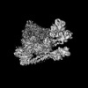 14098MC M: map data used to model this data C: citing same article ( |
|---|---|
| Similar structure data |

-Links
 PDBj
PDBj


- Assembly
| Deposited unit | 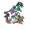
|
|---|---|
| 1 |
|
-Components
-Centromere protein ... , 14 types, 14 molecules CHIKLMNOPUQRTW
| #1: Protein | Mass: 61856.004 Da / Num. of mol.: 1 Source method: isolated from a genetically manipulated source Source: (gene. exp.) Homo sapiens (human) / Gene: CENPC, CENPC1, ICEN7 / Production host:  Trichoplusia ni (cabbage looper) / References: UniProt: Q03188 Trichoplusia ni (cabbage looper) / References: UniProt: Q03188 |
|---|---|
| #2: Protein | Mass: 28520.941 Da / Num. of mol.: 1 Source method: isolated from a genetically manipulated source Source: (gene. exp.) Homo sapiens (human) / Gene: CENPH, ICEN35 / Production host: Trichoplusia ni (cabbage looper) / Strain (production host): Tnao38 / References: UniProt: Q9H3R5 |
| #3: Protein | Mass: 86820.188 Da / Num. of mol.: 1 Source method: isolated from a genetically manipulated source Source: (gene. exp.) Homo sapiens (human) / Gene: CENPI, FSHPRH1, ICEN19, LRPR1 / Production host: Trichoplusia ni (cabbage looper) / Strain (production host): Tnao38 / References: UniProt: Q92674 |
| #4: Protein | Mass: 31696.070 Da / Num. of mol.: 1 Source method: isolated from a genetically manipulated source Source: (gene. exp.) Homo sapiens (human) / Gene: CENPK, ICEN37, FKSG14 / Production host: Trichoplusia ni (cabbage looper) / Strain (production host): Tnao38 / References: UniProt: Q9BS16 |
| #5: Protein | Mass: 39039.641 Da / Num. of mol.: 1 Source method: isolated from a genetically manipulated source Source: (gene. exp.) Homo sapiens (human) / Gene: CENPL, C1orf155, ICEN33 / Production host: Trichoplusia ni (cabbage looper) / Strain (production host): Tnao38 / References: UniProt: Q8N0S6 |
| #6: Protein | Mass: 19761.945 Da / Num. of mol.: 1 Source method: isolated from a genetically manipulated source Source: (gene. exp.) Homo sapiens (human) / Gene: CENPM, C22orf18, ICEN39, PANE1 / Production host: Trichoplusia ni (cabbage looper) / Strain (production host): Tnao38 / References: UniProt: Q9NSP4 |
| #7: Protein | Mass: 39609.551 Da / Num. of mol.: 1 Source method: isolated from a genetically manipulated source Source: (gene. exp.) Homo sapiens (human) / Gene: CENPN, C16orf60, ICEN32, BM-309 / Production host: Trichoplusia ni (cabbage looper) / Strain (production host): Tnao38 / References: UniProt: Q96H22 |
| #8: Protein | Mass: 33830.637 Da / Num. of mol.: 1 Source method: isolated from a genetically manipulated source Source: (gene. exp.) Homo sapiens (human) / Gene: CENPO, ICEN36, MCM21R / Production host: Trichoplusia ni (cabbage looper) / Strain (production host): Tnao38 / References: UniProt: Q9BU64 |
| #9: Protein | Mass: 33210.949 Da / Num. of mol.: 1 Source method: isolated from a genetically manipulated source Source: (gene. exp.) Homo sapiens (human) / Gene: CENPP / Production host: Trichoplusia ni (cabbage looper) / Strain (production host): Tnao38 / References: UniProt: Q6IPU0 |
| #10: Protein | Mass: 47609.766 Da / Num. of mol.: 1 Source method: isolated from a genetically manipulated source Source: (gene. exp.) Homo sapiens (human) / Gene: CENPU, ICEN24, KLIP1, MLF1IP, PBIP1 / Production host: Trichoplusia ni (cabbage looper) / Strain (production host): Tnao38 / References: UniProt: Q71F23 |
| #11: Protein | Mass: 30648.375 Da / Num. of mol.: 1 Source method: isolated from a genetically manipulated source Source: (gene. exp.) Homo sapiens (human) / Gene: CENPQ, C6orf139 / Production host: Trichoplusia ni (cabbage looper) / Strain (production host): Tnao38 / References: UniProt: Q7L2Z9 |
| #12: Protein | Mass: 20228.297 Da / Num. of mol.: 1 Source method: isolated from a genetically manipulated source Source: (gene. exp.) Homo sapiens (human) / Gene: ITGB3BP, CENPR, NRIF3 / Production host: Trichoplusia ni (cabbage looper) / Strain (production host): Tnao38 / References: UniProt: Q13352 |
| #13: Protein | Mass: 60502.613 Da / Num. of mol.: 1 Source method: isolated from a genetically manipulated source Source: (gene. exp.) Homo sapiens (human) / Gene: CENPT, C16orf56, ICEN22 / Plasmid: pETDuet / Production host:  |
| #14: Protein | Mass: 10087.236 Da / Num. of mol.: 1 Source method: isolated from a genetically manipulated source Source: (gene. exp.) Homo sapiens (human) / Gene: CENPW, C6orf173, CUG2 / Plasmid: pETDuet / Production host: |
-Protein/peptide , 1 types, 1 molecules X
| #15: Protein/peptide | Mass: 1294.587 Da / Num. of mol.: 1 Source method: isolated from a genetically manipulated source Source: (gene. exp.) Homo sapiens (human) / Production host: Trichoplusia ni (cabbage looper) / Strain (production host): Tnao38 |
|---|
-Experimental details
-Experiment
| Experiment | Method: ELECTRON MICROSCOPY |
|---|---|
| EM experiment | Aggregation state: PARTICLE / 3D reconstruction method: single particle reconstruction |
- Sample preparation
| Component | Name: human inner kinetochore CCAN complex / Type: COMPLEX / Entity ID: all / Source: MULTIPLE SOURCES |
|---|---|
| Molecular weight | Value: 0.451 MDa / Experimental value: NO |
| Source (natural) | Organism: Homo sapiens (human) |
| Source (recombinant) | Organism: Trichoplusia ni (cabbage looper) / Strain: Tnao38 |
| Buffer solution | pH: 6.8 / Details: 0.0025% Triton |
| Specimen | Conc.: 1.5 mg/ml / Embedding applied: NO / Shadowing applied: NO / Staining applied: NO / Vitrification applied: YES |
| Specimen support | Grid material: GOLD / Grid type: UltrAuFoil R1.2/1.3 |
| Vitrification | Instrument: FEI VITROBOT MARK IV / Cryogen name: ETHANE / Humidity: 100 % / Chamber temperature: 286 K / Details: blot for 3.5 seconds at blot force -3 |
- Electron microscopy imaging
| Experimental equipment |  Model: Titan Krios / Image courtesy: FEI Company |
|---|---|
| Microscopy | Model: FEI TITAN KRIOS |
| Electron gun | Electron source:  FIELD EMISSION GUN / Accelerating voltage: 300 kV / Illumination mode: OTHER FIELD EMISSION GUN / Accelerating voltage: 300 kV / Illumination mode: OTHER |
| Electron lens | Mode: BRIGHT FIELD / Nominal magnification: 130000 X / Nominal defocus max: 1200 nm / Nominal defocus min: 600 nm |
| Specimen holder | Cryogen: NITROGEN / Specimen holder model: FEI TITAN KRIOS AUTOGRID HOLDER |
| Image recording | Electron dose: 76.8 e/Å2 / Film or detector model: GATAN K3 (6k x 4k) / Num. of real images: 1540 Details: Images were collected in movie mode with 80 frames per image in superresolution mode with 0.35 A/px (0.7A native) |
| EM imaging optics | Energyfilter name: GIF Bioquantum / Energyfilter slit width: 20 eV / Phase plate: VOLTA PHASE PLATE |
- Processing
| EM software |
| ||||||||||||||||||||||||||||||||
|---|---|---|---|---|---|---|---|---|---|---|---|---|---|---|---|---|---|---|---|---|---|---|---|---|---|---|---|---|---|---|---|---|---|
| CTF correction | Type: PHASE FLIPPING AND AMPLITUDE CORRECTION | ||||||||||||||||||||||||||||||||
| Particle selection | Num. of particles selected: 140910 | ||||||||||||||||||||||||||||||||
| Symmetry | Point symmetry: C1 (asymmetric) | ||||||||||||||||||||||||||||||||
| 3D reconstruction | Resolution: 4.6 Å / Resolution method: FSC 0.143 CUT-OFF / Num. of particles: 25206 / Num. of class averages: 1 / Symmetry type: POINT | ||||||||||||||||||||||||||||||||
| Atomic model building | Protocol: AB INITIO MODEL / Space: REAL |
