 Movie
Movie Controller


+ Open data

- Basic information
| Entry | Database: PDB / ID: 7q5z | |||||||||||||||||||||||||||
|---|---|---|---|---|---|---|---|---|---|---|---|---|---|---|---|---|---|---|---|---|---|---|---|---|---|---|---|---|
| Title | Cryo-EM structure of native human A2ML1 | |||||||||||||||||||||||||||
 Components Components | Alpha-2-macroglobulin-like protein 1 | |||||||||||||||||||||||||||
 Keywords Keywords | IMMUNE SYSTEM / protease / inhibitor / thioester | |||||||||||||||||||||||||||
| Function / homology |  Function and homology information Function and homology informationregulation of endopeptidase activity / peptidase inhibitor activity / serine-type endopeptidase inhibitor activity / extracellular space / extracellular exosome Similarity search - Function | |||||||||||||||||||||||||||
| Biological species |  Homo sapiens (human) Homo sapiens (human) | |||||||||||||||||||||||||||
| Method | ELECTRON MICROSCOPY / single particle reconstruction / cryo EM / Resolution: 3.25 Å | |||||||||||||||||||||||||||
 Authors Authors | Zarantonello, A. / Nielsen, N.S. / Andersen, G.R. | |||||||||||||||||||||||||||
| Funding support |  Denmark, 1items Denmark, 1items
| |||||||||||||||||||||||||||
 Citation Citation | Journal: Nat Commun / Year: 2022 Title: Cryo-EM structures of human A2ML1 elucidate the protease-inhibitory mechanism of the A2M family. Authors: Nadia Sukusu Nielsen / Alessandra Zarantonello / Seandean Lykke Harwood / Kathrine Tejlgård Jensen / Katarzyna Kjøge / Ida B Thøgersen / Leif Schauser / Jesper Lykkegaard Karlsen / ...Authors: Nadia Sukusu Nielsen / Alessandra Zarantonello / Seandean Lykke Harwood / Kathrine Tejlgård Jensen / Katarzyna Kjøge / Ida B Thøgersen / Leif Schauser / Jesper Lykkegaard Karlsen / Gregers R Andersen / Jan J Enghild /  Abstract: A2ML1 is a monomeric protease inhibitor belonging to the A2M superfamily of protease inhibitors and complement factors. Here, we investigate the protease-inhibitory mechanism of human A2ML1 and ...A2ML1 is a monomeric protease inhibitor belonging to the A2M superfamily of protease inhibitors and complement factors. Here, we investigate the protease-inhibitory mechanism of human A2ML1 and determine the structures of its native and protease-cleaved conformations. The functional inhibitory unit of A2ML1 is a monomer that depends on covalent binding of the protease (mediated by A2ML1's thioester) to achieve inhibition. In contrast to the A2M tetramer which traps proteases in two internal chambers formed by four subunits, in protease-cleaved monomeric A2ML1 disordered regions surround the trapped protease and may prevent substrate access. In native A2ML1, the bait region is threaded through a hydrophobic channel, suggesting that disruption of this arrangement by bait region cleavage triggers the extensive conformational changes that result in protease inhibition. Structural comparisons with complement C3/C4 suggest that the A2M superfamily of proteins share this mechanism for the triggering of conformational change occurring upon proteolytic activation. | |||||||||||||||||||||||||||
| History |
|
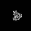
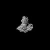
- Structure visualization
| Structure viewer | Molecule: MolmilJmol/JSmol |
|---|
- Downloads & links
-Download
| PDBx/mmCIF format | 7q5z.cif.gz | 248.1 KB | Display | PDBx/mmCIF format |
|---|---|---|---|---|
| PDB format | pdb7q5z.ent.gz | 192.7 KB | Display | PDB format |
| PDBx/mmJSON format | 7q5z.json.gz | Tree view | PDBx/mmJSON format | |
| Others |  Other downloads Other downloads |
-Validation report
| Summary document | 7q5z_validation.pdf.gz | 773.3 KB | Display | wwPDB validaton report |
|---|---|---|---|---|
| Full document | 7q5z_full_validation.pdf.gz | 782.1 KB | Display | |
| Data in XML | 7q5z_validation.xml.gz | 38.4 KB | Display | |
| Data in CIF | 7q5z_validation.cif.gz | 60.7 KB | Display | |
| Arichive directory | https://data.pdbj.org/pub/pdb/validation_reports/q5/7q5zftp://data.pdbj.org/pub/pdb/validation_reports/q5/7q5z | HTTPS FTP |
-Related structure data
| Related structure data |  13847MC  7q1yC  7q60C 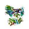 7q61C  7q62C M: map data used to model this data C: citing same article ( |
|---|---|
| Similar structure data |

-Links
 PDBj
PDBj

- Assembly
| Deposited unit | 
|
|---|---|
| 1 |
|
-Components
| #1: Protein | Mass: 159339.281 Da / Num. of mol.: 1 Source method: isolated from a genetically manipulated source Source: (gene. exp.) Homo sapiens (human) / Gene: A2ML1, CPAMD9 / Cell line (production host): HEK293F / Production host: Homo sapiens (human) / References: UniProt: A8K2U0 |
|---|---|
| Has ligand of interest | N |
| Has protein modification | Y |
-Experimental details
-Experiment
| Experiment | Method: ELECTRON MICROSCOPY |
|---|---|
| EM experiment | Aggregation state: PARTICLE / 3D reconstruction method: single particle reconstruction |
- Sample preparation
| Component | Name: Native A2ML1 / Type: COMPLEX / Entity ID: all / Source: RECOMBINANT |
|---|---|
| Source (natural) | Organism: Homo sapiens (human) |
| Source (recombinant) | Organism: Homo sapiens (human) / Strain: HEK293F |
| Buffer solution | pH: 7.2 |
| Specimen | Conc.: 1.2 mg/ml / Embedding applied: NO / Shadowing applied: NO / Staining applied: NO / Vitrification applied: YES |
| Vitrification | Cryogen name: PROPANE |
- Electron microscopy imaging
| Experimental equipment |  Model: Titan Krios / Image courtesy: FEI Company |
|---|---|
| Microscopy | Model: FEI TITAN KRIOS |
| Electron gun | Electron source:  FIELD EMISSION GUN / Accelerating voltage: 300 kV / Illumination mode: FLOOD BEAM FIELD EMISSION GUN / Accelerating voltage: 300 kV / Illumination mode: FLOOD BEAM |
| Electron lens | Mode: BRIGHT FIELD / Nominal defocus max: 1200 nm / Nominal defocus min: 600 nm |
| Image recording | Electron dose: 60 e/Å2 / Film or detector model: GATAN K3 BIOQUANTUM (6k x 4k) |
- Processing
| Software | Name: PHENIX / Version: 1.19.2_4158: / Classification: refinement | ||||||||||||||||||||||||
|---|---|---|---|---|---|---|---|---|---|---|---|---|---|---|---|---|---|---|---|---|---|---|---|---|---|
| EM software |
| ||||||||||||||||||||||||
| CTF correction | Details: cryoSPARC / Type: PHASE FLIPPING AND AMPLITUDE CORRECTION | ||||||||||||||||||||||||
| 3D reconstruction | Resolution: 3.25 Å / Resolution method: FSC 0.143 CUT-OFF / Num. of particles: 960516 / Symmetry type: POINT | ||||||||||||||||||||||||
| Refine LS restraints |
|
