[English] 日本語
Yorodumi
- PDB-7q1u: Structure of Hedgehog acyltransferase (HHAT) in complex with mega... -

+
Open data


ID or keywords:

Loading...

-
Basic information

Entry
Database: PDB / ID: 7q1u
TitleStructure of Hedgehog acyltransferase (HHAT) in complex with megabody 177 bound to non-hydrolysable palmitoyl-CoA (Composite Map)
Components
  • Megabody 177
  • Protein-cysteine N-palmitoyltransferase HHAT
KeywordsMEMBRANE PROTEIN / HHAT / inhibitor / palmitoyl-CoA / co enzyme A / Hedgehog acyl transferase / Sonic Hedgehog / SHH / MBOAT / morphogen / palmitoylation / signalling / endoplasmic reticulum / heme / small molecule binding / drug target
Function / homology
Function and homology information


N-terminal peptidyl-L-cysteine N-palmitoylation / O-acyltransferase activity / HHAT G278V doesn't palmitoylate Hh-Np / palmitoyltransferase activity / smoothened signaling pathway / Transferases; Acyltransferases; Transferring groups other than aminoacyl groups / Hedgehog ligand biogenesis / Golgi membrane / endoplasmic reticulum membrane / GTP binding ...N-terminal peptidyl-L-cysteine N-palmitoylation / O-acyltransferase activity / HHAT G278V doesn't palmitoylate Hh-Np / palmitoyltransferase activity / smoothened signaling pathway / Transferases; Acyltransferases; Transferring groups other than aminoacyl groups / Hedgehog ligand biogenesis / Golgi membrane / endoplasmic reticulum membrane / GTP binding / Golgi apparatus / endoplasmic reticulum
Similarity search - Function
Membrane bound O-acyl transferase, MBOAT / MBOAT, membrane-bound O-acyltransferase family
Similarity search - Domain/homology
CHOLESTEROL / Chem-HD6 / PROTOPORPHYRIN IX CONTAINING FE / PALMITIC ACID / Protein-cysteine N-palmitoyltransferase HHAT
Similarity search - Component
Biological speciesHomo sapiens (human)
Escherichia coli K-12 (bacteria)
MethodELECTRON MICROSCOPY / single particle reconstruction / cryo EM / Resolution: 2.7 Å
AuthorsCoupland, C. / Carrique, L. / Siebold, C.
Funding supportEuropean Union, 4items
OrganizationGrant numberCountry
Cancer Research UKC20724/A26752European Union
Cancer Research UKC20724/A14414European Union
Biotechnology and Biological Sciences Research Council (BBSRC)BB/T01508X/1European Union
European Research Council (ERC)647278European Union
CitationJournal: Mol Cell / Year: 2021
Title: Structure, mechanism, and inhibition of Hedgehog acyltransferase.
Authors: Claire E Coupland / Sebastian A Andrei / T Bertie Ansell / Loic Carrique / Pramod Kumar / Lea Sefer / Rebekka A Schwab / Eamon F X Byrne / Els Pardon / Jan Steyaert / Anthony I Magee / ...Authors: Claire E Coupland / Sebastian A Andrei / T Bertie Ansell / Loic Carrique / Pramod Kumar / Lea Sefer / Rebekka A Schwab / Eamon F X Byrne / Els Pardon / Jan Steyaert / Anthony I Magee / Thomas Lanyon-Hogg / Mark S P Sansom / Edward W Tate / Christian Siebold /
Abstract: The Sonic Hedgehog (SHH) morphogen pathway is fundamental for embryonic development and stem cell maintenance and is implicated in various cancers. A key step in signaling is transfer of a palmitate ...The Sonic Hedgehog (SHH) morphogen pathway is fundamental for embryonic development and stem cell maintenance and is implicated in various cancers. A key step in signaling is transfer of a palmitate group to the SHH N terminus, catalyzed by the multi-pass transmembrane enzyme Hedgehog acyltransferase (HHAT). We present the high-resolution cryo-EM structure of HHAT bound to substrate analog palmityl-coenzyme A and a SHH-mimetic megabody, revealing a heme group bound to HHAT that is essential for HHAT function. A structure of HHAT bound to potent small-molecule inhibitor IMP-1575 revealed conformational changes in the active site that occlude substrate binding. Our multidisciplinary analysis provides a detailed view of the mechanism by which HHAT adapts the membrane environment to transfer an acyl chain across the endoplasmic reticulum membrane. This structure of a membrane-bound O-acyltransferase (MBOAT) superfamily member provides a blueprint for other protein-substrate MBOATs and a template for future drug discovery.
History
DepositionOct 21, 2021Deposition site: PDBE / Processing site: PDBE
Revision 1.0Jun 8, 2022Provider: repository / Type: Initial release

-
Structure visualization

Structure viewerMolecule:
MolmilJmol/JSmol

Downloads & links

-
Assembly

Deposited unit
A: Protein-cysteine N-palmitoyltransferase HHAT
B: Megabody 177
hetero molecules


Theoretical massNumber of molelcules
Total (without water)163,7008
Polymers161,1682
Non-polymers2,5326
Water00
1


  • Idetical with deposited unit
  • defined by author
  • Evidence: electron microscopy
TypeNameSymmetry operationNumber
identity operation1_5551

-
Components

-
Protein / Antibody , 2 types, 2 molecules AB

#1: Protein Protein-cysteine N-palmitoyltransferase HHAT / Hedgehog acyltransferase / Melanoma antigen recognized by T-cells 2 / MART-2 / Skinny hedgehog protein 1


Mass: 58566.160 Da / Num. of mol.: 1
Source method: isolated from a genetically manipulated source
Source: (gene. exp.) Homo sapiens (human) / Gene: HHAT, MART2, SKI1 / Plasmid: pHR-CMV-TetO2 / Cell line (production host): HEK293S GnTI-/- / Production host: Homo sapiens (human)
References: UniProt: Q5VTY9, Transferases; Acyltransferases; Transferring groups other than aminoacyl groups
#2: Antibody Megabody 177


Mass: 102601.586 Da / Num. of mol.: 1
Source method: isolated from a genetically manipulated source
Source: (gene. exp.) Escherichia coli K-12 (bacteria) / Plasmid: pMESP23E2 / Production host: Escherichia coli (E. coli) / Strain (production host): WK6

-
Non-polymers , 5 types, 6 molecules

#3: Chemical ChemComp-CLR / CHOLESTEROL


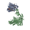
Mass: 386.654 Da / Num. of mol.: 1 / Source method: obtained synthetically / Formula: C27H46O
#4: Chemical ChemComp-HEM / PROTOPORPHYRIN IX CONTAINING FE / HEME


Mass: 616.487 Da / Num. of mol.: 1 / Source method: obtained synthetically / Formula: C34H32FeN4O4 / Feature type: SUBJECT OF INVESTIGATION
#5: Chemical ChemComp-HD6 / [[(2~{R},3~{S},4~{R},5~{R})-5-(6-aminopurin-9-yl)-4-oxidanyl-3-phosphonooxy-oxolan-2-yl]methoxy-oxidanyl-phosphoryl] [(3~{S})-4-[[3-(2-hexadecylsulfanylethylamino)-3-oxidanylidene-propyl]amino]-2,2-dimethyl-3-oxidanyl-4-oxidanylidene-butyl] hydrogen phosphate


Mass: 991.959 Da / Num. of mol.: 1 / Source method: obtained synthetically / Formula: C37H68N7O16P3S / Feature type: SUBJECT OF INVESTIGATION
#6: Chemical ChemComp-MG / MAGNESIUM ION


Mass: 24.305 Da / Num. of mol.: 1 / Source method: obtained synthetically / Formula: Mg
#7: Chemical ChemComp-PLM / PALMITIC ACID


Mass: 256.424 Da / Num. of mol.: 2 / Source method: obtained synthetically / Formula: C16H32O2

-
Details

Has ligand of interestY

-
Experimental details

-
Experiment

ExperimentMethod: ELECTRON MICROSCOPY
EM experimentAggregation state: PARTICLE / 3D reconstruction method: single particle reconstruction

-
Sample preparation

Component
IDNameTypeEntity IDParent-IDSource
1Hedgehog acyltransferase (HHAT) bound to non hydrolysable palmitoyl-CoA in complex with megabody 177COMPLEX#1-#20MULTIPLE SOURCES
2Megabody 177COMPLEX#21RECOMBINANT
3Hedgehog acyltransferase (HHAT) bound to non hydrolysable palmitoyl-CoACOMPLEX#11RECOMBINANT
Molecular weight
IDEntity assembly-IDExperimental value
11NO
21NO
31NO
Source (natural)
IDEntity assembly-IDOrganismNcbi tax-ID
32Escherichia coli K-12 (bacteria)83333
43Homo sapiens (human)9606
Source (recombinant)
IDEntity assembly-IDOrganismNcbi tax-IDStrainCellPlasmid
32Escherichia coli (E. coli)562WK6pMESP23E2
43Homo sapiens (human)9606HEK293S GnTI-/-pHR-CMV-TetO2
Buffer solutionpH: 8
Buffer component
IDConc.NameFormulaBuffer-ID
120 mMTris-HCl1
2150 mMsodium chlorideNaCl1
35 mM16:0 Ether Coenzyme AC37H77N10O16P3S1
SpecimenConc.: 4 mg/ml / Embedding applied: NO / Shadowing applied: NO / Staining applied: NO / Vitrification applied: YES
Specimen supportGrid material: COPPER / Grid mesh size: 300 divisions/in. / Grid type: Quantifoil R1.2/1.3
VitrificationInstrument: FEI VITROBOT MARK IV / Cryogen name: ETHANE / Humidity: 100 % / Chamber temperature: 293 K

-
Electron microscopy imaging

Experimental equipment
Model: Titan Krios / Image courtesy: FEI Company
MicroscopyModel: FEI TITAN KRIOS
Electron gunElectron source: FIELD EMISSION GUN / Accelerating voltage: 300 kV / Illumination mode: FLOOD BEAM
Electron lensMode: BRIGHT FIELD / Nominal defocus max: 2500 nm / Nominal defocus min: 1000 nm / Cs: 2.7 mm / C2 aperture diameter: 70 µm / Alignment procedure: COMA FREE
Specimen holderCryogen: NITROGEN / Specimen holder model: FEI TITAN KRIOS AUTOGRID HOLDER
Image recordingElectron dose: 54.8 e/Å2 / Film or detector model: GATAN K3 BIOQUANTUM (6k x 4k) / Num. of grids imaged: 1 / Num. of real images: 3637
EM imaging opticsEnergyfilter name: GIF Bioquantum / Energyfilter slit width: 20 eV

-
Processing

Software
NameVersionClassification
phenix.real_space_refine1.19.2_4158refinement
PHENIX1.19.2_4158refinement
EM software
IDNameVersionCategory
2EPUimage acquisition
4cryoSPARCV3.1.1CTF correction
7UCSF ChimeraX1.2model fitting
9cryoSPARCV3.1.1initial Euler assignment
10cryoSPARCV3.1.1final Euler assignment
11RELION3.1.1classification
12cryoSPARCV3.1.13D reconstruction
13PHENIX1.19.2model refinement
14Coot0.9.2model refinement
CTF correctionType: PHASE FLIPPING AND AMPLITUDE CORRECTION
Particle selectionNum. of particles selected: 977000
SymmetryPoint symmetry: C1 (asymmetric)
3D reconstructionResolution: 2.7 Å / Resolution method: FSC 0.143 CUT-OFF / Num. of particles: 238000 / Num. of class averages: 1 / Symmetry type: POINT
Atomic model buildingB value: 56 / Protocol: AB INITIO MODEL / Space: REAL
RefinementCross valid method: NONE
Stereochemistry target values: GeoStd + Monomer Library + CDL v1.2
Displacement parametersBiso mean: 50.91 Å2
Refine LS restraints
Refine-IDTypeDev idealNumber
ELECTRON MICROSCOPYf_bond_d0.003211444
ELECTRON MICROSCOPYf_angle_d0.484515575
ELECTRON MICROSCOPYf_chiral_restr0.03851636
ELECTRON MICROSCOPYf_plane_restr0.00311956
ELECTRON MICROSCOPYf_dihedral_angle_d5.82741621

+
About Yorodumi

-
News

-
Feb 9, 2022. New format data for meta-information of EMDB entries

New format data for meta-information of EMDB entries

  • Version 3 of the EMDB header file is now the official format.
  • The previous official version 1.9 will be removed from the archive.

Related info.:EMDB header

External links:wwPDB to switch to version 3 of the EMDB data model

-
Aug 12, 2020. Covid-19 info

Covid-19 info

URL: https://pdbjlvh1.pdbj.org/emnavi/covid19.php

New page: Covid-19 featured information page in EM Navigator.

Related info.:Covid-19 info / Mar 5, 2020. Novel coronavirus structure data

+
Mar 5, 2020. Novel coronavirus structure data

Novel coronavirus structure data

Related info.:Yorodumi Speices / Aug 12, 2020. Covid-19 info

External links:COVID-19 featured content - PDBj / Molecule of the Month (242):Coronavirus Proteases

+
Jan 31, 2019. EMDB accession codes are about to change! (news from PDBe EMDB page)

EMDB accession codes are about to change! (news from PDBe EMDB page)

  • The allocation of 4 digits for EMDB accession codes will soon come to an end. Whilst these codes will remain in use, new EMDB accession codes will include an additional digit and will expand incrementally as the available range of codes is exhausted. The current 4-digit format prefixed with “EMD-” (i.e. EMD-XXXX) will advance to a 5-digit format (i.e. EMD-XXXXX), and so on. It is currently estimated that the 4-digit codes will be depleted around Spring 2019, at which point the 5-digit format will come into force.
  • The EM Navigator/Yorodumi systems omit the EMD- prefix.

Related info.:Q: What is EMD? / ID/Accession-code notation in Yorodumi/EM Navigator

External links:EMDB Accession Codes are Changing Soon! / Contact to PDBj

+
Jul 12, 2017. Major update of PDB

Major update of PDB

  • wwPDB released updated PDB data conforming to the new PDBx/mmCIF dictionary.
  • This is a major update changing the version number from 4 to 5, and with Remediation, in which all the entries are updated.
  • In this update, many items about electron microscopy experimental information are reorganized (e.g. em_software).
  • Now, EM Navigator and Yorodumi are based on the updated data.

External links:wwPDB Remediation / Enriched Model Files Conforming to OneDep Data Standards Now Available in the PDB FTP Archive

-
Yorodumi

Thousand views of thousand structures

  • Yorodumi is a browser for structure data from EMDB, PDB, SASBDB, etc.
  • This page is also the successor to EM Navigator detail page, and also detail information page/front-end page for Omokage search.
  • The word "yorodu" (or yorozu) is an old Japanese word meaning "ten thousand". "mi" (miru) is to see.

Related info.:EMDB / PDB / SASBDB / Comparison of 3 databanks / Yorodumi Search / Aug 31, 2016. New EM Navigator & Yorodumi / Yorodumi Papers / Jmol/JSmol / Function and homology information / Changes in new EM Navigator and Yorodumi

Read more