 Movie
Movie Controller

[English] 日本語
 Yorodumi
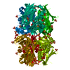
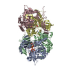
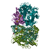
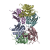
- PDB-7pq5: Photorhabdus laumondii T6SS-associated Rhs protein carrying the T... -
+ Open data

- Basic information
| Entry | Database: PDB / ID: 7pq5 | ||||||
|---|---|---|---|---|---|---|---|
| Title | Photorhabdus laumondii T6SS-associated Rhs protein carrying the Tre23 toxin domain | ||||||
 Components Components | Tre23 | ||||||
 Keywords Keywords | TOXIN / RHS / TVISS / T6SS | ||||||
| Function / homology |  Function and homology information Function and homology informationRHS repeat / RHS Repeat / Domain of unknown function DUF6531 / Domain of unknown function (DUF6531) / PAAR motif / PAAR motif / : / Teneurin YD-shell / YD repeat / : / Rhs repeat-associated core Similarity search - Domain/homology | ||||||
| Biological species |  Photorhabdus laumondii subsp. laumondii TTO1 (bacteria) Photorhabdus laumondii subsp. laumondii TTO1 (bacteria) | ||||||
| Method | ELECTRON MICROSCOPY / single particle reconstruction / cryo EM / Resolution: 3.17 Å | ||||||
 Authors Authors | Jurenas, D. / Talachia Rosa, L. / Rey, M. / Chamot-Rooke, J. / Fronzes, R. / Cascales, E. | ||||||
| Funding support |  France, 1items France, 1items
| ||||||
 Citation Citation | Journal: Nat Commun / Year: 2021 Title: Mounting, structure and autocleavage of a type VI secretion-associated Rhs polymorphic toxin. Authors: Dukas Jurėnas / Leonardo Talachia Rosa / Martial Rey / Julia Chamot-Rooke / Rémi Fronzes / Eric Cascales / Abstract: Bacteria have evolved toxins to outcompete other bacteria or to hijack host cell pathways. One broad family of bacterial polymorphic toxins gathers multidomain proteins with a modular organization, ...Bacteria have evolved toxins to outcompete other bacteria or to hijack host cell pathways. One broad family of bacterial polymorphic toxins gathers multidomain proteins with a modular organization, comprising a C-terminal toxin domain fused to a N-terminal domain that adapts to the delivery apparatus. Polymorphic toxins include bacteriocins, contact-dependent growth inhibition systems, and specialized Hcp, VgrG, PAAR or Rhs Type VI secretion (T6SS) components. We recently described and characterized Tre23, a toxin domain fused to a T6SS-associated Rhs protein in Photorhabdus laumondii, Rhs1. Here, we show that Rhs1 forms a complex with the T6SS spike protein VgrG and the EagR chaperone. Using truncation derivatives and cross-linking mass spectrometry, we demonstrate that VgrG-EagR-Rhs1 complex formation requires the VgrG C-terminal β-helix and the Rhs1 N-terminal region. We then report the cryo-electron-microscopy structure of the Rhs1-EagR complex, demonstrating that the Rhs1 central region forms a β-barrel cage-like structure that encapsulates the C-terminal toxin domain, and provide evidence for processing of the Rhs1 protein through aspartyl autoproteolysis. We propose a model for Rhs1 loading on the T6SS, transport and delivery into the target cell. | ||||||
| History |
|
- Structure visualization
| Movie |
Movie viewer |
|---|---|
| Structure viewer | Molecule: MolmilJmol/JSmol |
- Downloads & links
-Download
| PDBx/mmCIF format | 7pq5.cif.gz | 209.4 KB | Display | PDBx/mmCIF format |
|---|---|---|---|---|
| PDB format | pdb7pq5.ent.gz | 156.4 KB | Display | PDB format |
| PDBx/mmJSON format | 7pq5.json.gz | Tree view | PDBx/mmJSON format | |
| Others |  Other downloads Other downloads |
-Validation report
| Arichive directory | https://data.pdbj.org/pub/pdb/validation_reports/pq/7pq5ftp://data.pdbj.org/pub/pdb/validation_reports/pq/7pq5 | HTTPS FTP |
|---|
-Related structure data
| Related structure data |  13587MC M: map data used to model this data C: citing same article ( |
|---|---|
| Similar structure data |


-Links
 PDBj
PDBj- Assembly
| Deposited unit | 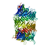
|
|---|---|
| 1 |
|
-Components
| #1: Protein | Mass: 165588.828 Da / Num. of mol.: 1 / Mutation: D1338N Source method: isolated from a genetically manipulated source Source: (gene. exp.) Photorhabdus laumondii subsp. laumondii TTO1 (bacteria)Gene: plu0353 / Production host: |
|---|
-Experimental details
-Experiment
| Experiment | Method: ELECTRON MICROSCOPY |
|---|---|
| EM experiment | Aggregation state: PARTICLE / 3D reconstruction method: single particle reconstruction |
- Sample preparation
| Component | Name: Tre23 / Type: COMPLEX / Entity ID: all / Source: RECOMBINANT |
|---|---|
| Molecular weight | Experimental value: NO |
| Source (natural) | Organism: Photorhabdus laumondii (bacteria) |
| Source (recombinant) | Organism: |
| Buffer solution | pH: 8.5 Details: 50 mM Tris-HCl pH8.5, 250 mM NaCl, 1 mM TCEP, cOmpleteTM protease inhibitor cocktail (Sigma-Aldrich) |
| Specimen | Conc.: 0.5 mg/ml / Embedding applied: NO / Shadowing applied: NO / Staining applied: NO / Vitrification applied: YES |
| Specimen support | Details: 2 mAu current / Grid material: COPPER / Grid mesh size: 200 divisions/in. / Grid type: Quantifoil R2/2 |
| Vitrification | Instrument: FEI VITROBOT MARK III / Cryogen name: ETHANE / Humidity: 100 % / Chamber temperature: 277 K |
- Electron microscopy imaging
| Experimental equipment |  Model: Talos Arctica / Image courtesy: FEI Company |
|---|---|
| Microscopy | Model: FEI TALOS ARCTICA |
| Electron gun | Electron source:  FIELD EMISSION GUN / Accelerating voltage: 200 kV / Illumination mode: FLOOD BEAM FIELD EMISSION GUN / Accelerating voltage: 200 kV / Illumination mode: FLOOD BEAM |
| Electron lens | Mode: BRIGHT FIELD / Nominal magnification: 45000 X / Nominal defocus max: 20000 nm / Nominal defocus min: 5000 nm / Cs: 2.7 mm |
| Specimen holder | Cryogen: NITROGEN |
| Image recording | Average exposure time: 3.7 sec. / Electron dose: 49.75 e/Å2 / Detector mode: COUNTING / Film or detector model: GATAN K2 SUMMIT (4k x 4k) / Num. of grids imaged: 1 / Num. of real images: 8550 |
| Image scans | Movie frames/image: 37 / Used frames/image: 1-37 |
- Processing
| Software |
| ||||||||||||||||||||||||||||||||||||
|---|---|---|---|---|---|---|---|---|---|---|---|---|---|---|---|---|---|---|---|---|---|---|---|---|---|---|---|---|---|---|---|---|---|---|---|---|---|
| EM software |
| ||||||||||||||||||||||||||||||||||||
| CTF correction | Type: PHASE FLIPPING AND AMPLITUDE CORRECTION | ||||||||||||||||||||||||||||||||||||
| Particle selection | Num. of particles selected: 2623687 | ||||||||||||||||||||||||||||||||||||
| 3D reconstruction | Resolution: 3.17 Å / Resolution method: FSC 0.143 CUT-OFF / Num. of particles: 140577 / Num. of class averages: 1 / Symmetry type: POINT | ||||||||||||||||||||||||||||||||||||
| Atomic model building | B value: 75.76 / Protocol: BACKBONE TRACE / Space: REAL | ||||||||||||||||||||||||||||||||||||
| Refinement | Highest resolution: 3.17 Å / Cross valid method: NONE Stereochemistry target values: GeoStd + Monomer Library + CDL v1.2 | ||||||||||||||||||||||||||||||||||||
| Displacement parameters | Biso mean: 47.88 Å2 | ||||||||||||||||||||||||||||||||||||
| Refine LS restraints |
|
