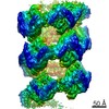
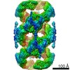
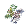
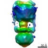
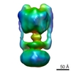
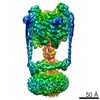
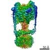
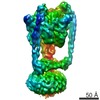
ジャーナル: Proc Natl Acad Sci U S A / 年: 2022 タイトル: Helical self-assembly of a mucin segment suggests an evolutionary origin for von Willebrand factor tubules. 著者: Gabriel Javitt / Deborah Fass / 要旨: The glycoprotein von Willebrand factor (VWF) contributes to hemostasis by stanching injuries in blood vessel walls. A distinctive feature of VWF is its assembly into long, helical tubules in ...The glycoprotein von Willebrand factor (VWF) contributes to hemostasis by stanching injuries in blood vessel walls. A distinctive feature of VWF is its assembly into long, helical tubules in endothelial cells prior to secretion. When VWF is released into the bloodstream, these tubules unfurl to release linear polymers that bind subendothelial collagen at wound sites, recruit platelets, and initiate the clotting cascade. VWF evolved from gel-forming mucins, the polymeric glycoproteins that coat and protect exposed epithelia. Despite the divergent function of VWF in blood vessel repair, sequence conservation and shared domain organization imply that VWF retained key aspects of the mucin bioassembly mechanism. Here, we show using cryo-electron microscopy that the ability to form tubules, a property hitherto thought to have arisen as a VWF adaptation to the vasculature, is a feature of the amino-terminal region of mucin. This segment of the human intestinal gel-forming mucin (MUC2) was found to self-assemble into tubules with a striking resemblance to those of VWF itself. To facilitate a comparison, we determined the residue-resolution structure of tubules formed by the homologous segment of VWF. The structures of the MUC2 and VWF tubules revealed the flexible joints and the intermolecular interactions required for tubule formation. Steric constraints in full-length MUC2 suggest that linear filaments, a previously observed supramolecular assembly form, are more likely than tubules to be the physiological mucin storage intermediate. Nevertheless, MUC2 tubules indicate a possible evolutionary origin for VWF tubules and elucidate design principles present in mucins and VWF.
履歴
登録
2021年9月13日
登録サイト: PDBE / 処理サイト: PDBE
改定 1.0
2022年2月16日
Provider: repository / タイプ: Initial release
改定 1.0
2022年2月16日
Data content type: EM metadata / Data content type: EM metadata / Provider: repository / タイプ: Initial release
改定 1.0
2022年2月16日
Data content type: FSC / Data content type: FSC / Provider: repository / タイプ: Initial release
改定 1.0
2022年2月16日
Data content type: Image / Data content type: Image / Provider: repository / タイプ: Initial release
改定 1.0
2022年2月16日
Data content type: Primary map / Data content type: Primary map / Provider: repository / タイプ: Initial release
改定 1.0
2022年2月16日
Data content type: FSC / Data content type: FSC / Provider: repository / タイプ: Initial release
改定 1.0
2022年2月16日
Data content type: Image / Data content type: Image / Provider: repository / タイプ: Initial release
改定 1.0
2022年2月16日
Data content type: Primary map / Data content type: Primary map / Provider: repository / タイプ: Initial release
Data content type: EM metadata / Data content type: EM metadata / EM metadata / Group: Data processing / Experimental summary / Data content type: EM metadata / EM metadata / カテゴリ: em_admin / em_software / Data content type: EM metadata / EM metadata / Item: _em_admin.last_update / _em_software.name
 ムービー
ムービー コントローラー


 データを開く
 基本情報
基本情報 要素
要素 キーワード
キーワード 機能・相同性情報
 Homo sapiens (ヒト)
 データ登録者
 引用
引用
 構造の表示
構造の表示 ダウンロードとリンク
 その他のダウンロード
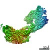


 PDBj
PDBj 集合体
集合体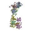

 分子量: 40.078 Da / 分子数: 12 / 由来タイプ: 合成 / 式: Ca
 タイプ: D-saccharide, beta linking / 分子量: 221.208 Da / 分子数: 8 / 由来タイプ: 合成 / 式: C8H15NO6
 分子量: 18.015 Da / 分子数: 92 / 由来タイプ: 天然 / 式: H2O
 試料調製
試料調製 電子顕微鏡撮影
 FIELD EMISSION GUN / 加速電圧: 300 kV / 照射モード: FLOOD BEAM
 解析
解析