 Movie
Movie Controller


+ Open data

- Basic information
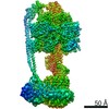
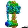
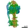
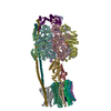
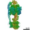
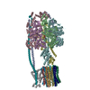
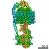
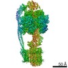
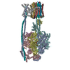
| Entry | Database: PDB / ID: 7p3w | ||||||
|---|---|---|---|---|---|---|---|
| Title | F1Fo-ATP synthase from Acinetobacter baumannii (state 3) | ||||||
 Components Components | (ATP synthase ...) x 8 | ||||||
 Keywords Keywords | MEMBRANE PROTEIN / ATP synthase / ESKAPE / Rotary ATP synthase / F1Fo / peptidisc / bioenergetics / IMP / multi-drug resistance / pathogenic | ||||||
| Function / homology |  Function and homology information Function and homology informationproton motive force-driven plasma membrane ATP synthesis / proton-transporting two-sector ATPase complex, proton-transporting domain / proton-transporting ATPase activity, rotational mechanism / H+-transporting two-sector ATPase / proton-transporting ATP synthase complex / proton-transporting ATP synthase activity, rotational mechanism / ADP binding / lipid binding / ATP binding / plasma membrane Similarity search - Function | ||||||
| Biological species |  Acinetobacter baumannii (bacteria) Acinetobacter baumannii (bacteria) | ||||||
| Method | ELECTRON MICROSCOPY / single particle reconstruction / cryo EM / Resolution: 4.3 Å | ||||||
 Authors Authors | Demmer, J.K. / Phillips, B.P. / Uhrig, O.L. / Filloux, A. / Allsopp, L.P. / Bublitz, M. / Meier, T. | ||||||
| Funding support |  United Kingdom, 1items United Kingdom, 1items
| ||||||
 Citation Citation | Journal: Sci Adv / Year: 2022 Title: Structure of ATP synthase from ESKAPE pathogen . Authors: Julius K Demmer / Ben P Phillips / O Lisa Uhrig / Alain Filloux / Luke P Allsopp / Maike Bublitz / Thomas Meier / Abstract: The global spread of multidrug-resistant infections urgently calls for the identification of novel drug targets. We solved the electron cryo-microscopy structure of the FF-adenosine 5'-triphosphate ...The global spread of multidrug-resistant infections urgently calls for the identification of novel drug targets. We solved the electron cryo-microscopy structure of the FF-adenosine 5'-triphosphate (ATP) synthase from in three distinct conformational states. The nucleotide-converting F subcomplex reveals a specific self-inhibition mechanism, which supports a unidirectional ratchet mechanism to avoid wasteful ATP consumption. In the membrane-embedded F complex, the structure shows unique structural adaptations along both the entry and exit pathways of the proton-conducting a-subunit. These features, absent in mitochondrial ATP synthases, represent attractive targets for the development of next-generation therapeutics that can act directly at the culmination of bioenergetics in this clinically relevant pathogen. | ||||||
| History |
|
- Structure visualization
| Movie |
Movie viewer |
|---|---|
| Structure viewer | Molecule: MolmilJmol/JSmol |
- Downloads & links
-Download
| PDBx/mmCIF format | 7p3w.cif.gz | 802.8 KB | Display | PDBx/mmCIF format |
|---|---|---|---|---|
| PDB format | pdb7p3w.ent.gz | 669.6 KB | Display | PDB format |
| PDBx/mmJSON format | 7p3w.json.gz | Tree view | PDBx/mmJSON format | |
| Others |  Other downloads Other downloads |
-Validation report
| Arichive directory | https://data.pdbj.org/pub/pdb/validation_reports/p3/7p3wftp://data.pdbj.org/pub/pdb/validation_reports/p3/7p3w | HTTPS FTP |
|---|
-Related structure data
| Related structure data | 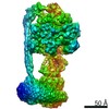 13186MC 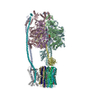 7p2yC 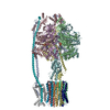 7p3nC C: citing same article ( M: map data used to model this data |
|---|---|
| Similar structure data |


-Links
 PDBj
PDBj


- Assembly
| Deposited unit | 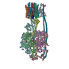
|
|---|---|
| 1 |
|
-Components
-ATP synthase ... , 8 types, 22 molecules ABCDEFGHJKLOPQRSabpdeg
| #1: Protein | Mass: 55452.906 Da / Num. of mol.: 3 / Source method: isolated from a natural source Source: (natural) Acinetobacter baumannii (strain ATCC 17978 / CIP 53.77 / LMG 1025 / NCDC KC755 / 5377) (bacteria)Strain: ATCC 17978 / CIP 53.77 / LMG 1025 / NCDC KC755 / 5377 References: UniProt: A3M142, H+-transporting two-sector ATPase #2: Protein | Mass: 50327.180 Da / Num. of mol.: 3 / Source method: isolated from a natural source Source: (natural) Acinetobacter baumannii (strain ATCC 17978 / CIP 53.77 / LMG 1025 / NCDC KC755 / 5377) (bacteria)Strain: ATCC 17978 / CIP 53.77 / LMG 1025 / NCDC KC755 / 5377 References: UniProt: A3M144, H+-transporting two-sector ATPase #3: Protein | Mass: 8363.021 Da / Num. of mol.: 10 / Source method: isolated from a natural source Source: (natural) Acinetobacter baumannii (strain ATCC 17978 / CIP 53.77 / LMG 1025 / NCDC KC755 / 5377) (bacteria)Strain: ATCC 17978 / CIP 53.77 / LMG 1025 / NCDC KC755 / 5377 References: UniProt: A3M139 #4: Protein | | Mass: 32467.396 Da / Num. of mol.: 1 / Source method: isolated from a natural source Source: (natural) Acinetobacter baumannii (strain ATCC 17978 / CIP 53.77 / LMG 1025 / NCDC KC755 / 5377) (bacteria)Strain: ATCC 17978 / CIP 53.77 / LMG 1025 / NCDC KC755 / 5377 References: UniProt: A3M137 #5: Protein | Mass: 17009.312 Da / Num. of mol.: 2 / Source method: isolated from a natural source Source: (natural) Acinetobacter baumannii (strain ATCC 17978 / CIP 53.77 / LMG 1025 / NCDC KC755 / 5377) (bacteria)Strain: ATCC 17978 / CIP 53.77 / LMG 1025 / NCDC KC755 / 5377 References: UniProt: A3M140 #6: Protein | | Mass: 19525.010 Da / Num. of mol.: 1 / Source method: isolated from a natural source Source: (natural) Acinetobacter baumannii (strain ATCC 17978 / CIP 53.77 / LMG 1025 / NCDC KC755 / 5377) (bacteria)Strain: ATCC 17978 / CIP 53.77 / LMG 1025 / NCDC KC755 / 5377 References: UniProt: A3M141 #7: Protein | | Mass: 14551.682 Da / Num. of mol.: 1 / Source method: isolated from a natural source Source: (natural) Acinetobacter baumannii (strain ATCC 17978 / CIP 53.77 / LMG 1025 / NCDC KC755 / 5377) (bacteria)Strain: ATCC 17978 / CIP 53.77 / LMG 1025 / NCDC KC755 / 5377 References: UniProt: A3M145 #8: Protein | | Mass: 32135.037 Da / Num. of mol.: 1 / Source method: isolated from a natural source Source: (natural) Acinetobacter baumannii (strain ATCC 17978 / CIP 53.77 / LMG 1025 / NCDC KC755 / 5377) (bacteria)Strain: ATCC 17978 / CIP 53.77 / LMG 1025 / NCDC KC755 / 5377 References: UniProt: A3M143 |
|---|
-Non-polymers , 3 types, 8 molecules 


| #9: Chemical |  Mass: 507.181 Da / Num. of mol.: 3 / Source method: obtained synthetically / Formula: C10H16N5O13P3 / Comment: ATP, energy-carrying molecule*YM Mass: 507.181 Da / Num. of mol.: 3 / Source method: obtained synthetically / Formula: C10H16N5O13P3 / Comment: ATP, energy-carrying molecule*YM#10: Chemical | ChemComp-MG /  Mass: 24.305 Da / Num. of mol.: 4 / Source method: obtained synthetically / Formula: Mg Mass: 24.305 Da / Num. of mol.: 4 / Source method: obtained synthetically / Formula: Mg#11: Chemical | ChemComp-ADP / |  Mass: 427.201 Da / Num. of mol.: 1 / Source method: obtained synthetically / Formula: C10H15N5O10P2 / Comment: ADP, energy-carrying molecule*YM Mass: 427.201 Da / Num. of mol.: 1 / Source method: obtained synthetically / Formula: C10H15N5O10P2 / Comment: ADP, energy-carrying molecule*YM |
|---|
-Details
| Has ligand of interest | N |
|---|
-Experimental details
-Experiment
| Experiment | Method: ELECTRON MICROSCOPY |
|---|---|
| EM experiment | Aggregation state: PARTICLE / 3D reconstruction method: single particle reconstruction |
- Sample preparation
| Component | Name: F1Fo ATP synthase / Type: COMPLEX Details: State 1 of F1Fo ATP synthase from ESKAPE pathogen Acinetobacter baumannii Entity ID: #1-#8 / Source: NATURAL | ||||||||||||||||||||
|---|---|---|---|---|---|---|---|---|---|---|---|---|---|---|---|---|---|---|---|---|---|
| Molecular weight | Value: 0.528 MDa / Experimental value: NO | ||||||||||||||||||||
| Source (natural) | Organism: Acinetobacter baumannii ATCC 17978 (bacteria) / Cellular location: Cell membrane | ||||||||||||||||||||
| Buffer solution | pH: 6.8 | ||||||||||||||||||||
| Buffer component |
| ||||||||||||||||||||
| Specimen | Conc.: 0.05 mg/ml / Embedding applied: NO / Shadowing applied: NO / Staining applied: NO / Vitrification applied: YES Details: Sample was applied directly from gel filtration to ultra-thin carbon in peptidiscs. | ||||||||||||||||||||
| Specimen support | Grid material: COPPER / Grid mesh size: 200 divisions/in. / Grid type: PELCO Ultrathin Carbon with Lacey Carbon | ||||||||||||||||||||
| Vitrification | Instrument: FEI VITROBOT MARK IV / Cryogen name: ETHANE / Humidity: 100 % / Chamber temperature: 281 K / Details: 4ul sample, blotted for 4s at a blotforce of -4. |
- Electron microscopy imaging
| Experimental equipment |  Model: Titan Krios / Image courtesy: FEI Company |
|---|---|
| Microscopy | Model: FEI TITAN KRIOS Details: Data collected at Diamond Light Source (Harwell, UK) using Titan Krios G3 and automated alignments using Sherpa. Detector used was Gatan K3 including energy filter. |
| Electron gun | Electron source:  FIELD EMISSION GUN / Accelerating voltage: 300 kV / Illumination mode: FLOOD BEAM FIELD EMISSION GUN / Accelerating voltage: 300 kV / Illumination mode: FLOOD BEAM |
| Electron lens | Mode: BRIGHT FIELD / Nominal magnification: 85000 X / Nominal defocus max: 2500 nm / Nominal defocus min: 1250 nm / Cs: 2.7 mm / C2 aperture diameter: 100 µm / Alignment procedure: BASIC |
| Specimen holder | Cryogen: NITROGEN / Specimen holder model: FEI TITAN KRIOS AUTOGRID HOLDER |
| Image recording | Electron dose: 60 e/Å2 / Film or detector model: GATAN K3 (6k x 4k) / Num. of grids imaged: 1 / Num. of real images: 11490 |
| EM imaging optics | Energyfilter slit width: 20 eV |
| Image scans | Width: 5760 / Height: 4092 |
- Processing
| Software | Name: UCSF ChimeraX / Version: 1.1/v9 / Classification: model building / URL: https://www.rbvi.ucsf.edu/chimerax/ / Os: Windows / Type: package | ||||||||||||||||||||||||||||
|---|---|---|---|---|---|---|---|---|---|---|---|---|---|---|---|---|---|---|---|---|---|---|---|---|---|---|---|---|---|
| EM software |
| ||||||||||||||||||||||||||||
| CTF correction | Type: PHASE FLIPPING AND AMPLITUDE CORRECTION | ||||||||||||||||||||||||||||
| Particle selection | Num. of particles selected: 349160 | ||||||||||||||||||||||||||||
| 3D reconstruction | Resolution: 4.3 Å / Resolution method: FSC 0.143 CUT-OFF / Num. of particles: 25428 / Num. of class averages: 1 / Symmetry type: POINT |
