 Movie
Movie Controller


+ Open data

- Basic information
| Entry | Database: PDB / ID: 7p1t | ||||||||||||||||||||||||||||||||||||||||||||||||||||||||||||||||||||||||
|---|---|---|---|---|---|---|---|---|---|---|---|---|---|---|---|---|---|---|---|---|---|---|---|---|---|---|---|---|---|---|---|---|---|---|---|---|---|---|---|---|---|---|---|---|---|---|---|---|---|---|---|---|---|---|---|---|---|---|---|---|---|---|---|---|---|---|---|---|---|---|---|---|---|
| Title | Cryo-EM structure of encapsulin from Mycobacterium tuberculosis | ||||||||||||||||||||||||||||||||||||||||||||||||||||||||||||||||||||||||
 Components Components | 29 kDa antigen, Cfp29 | ||||||||||||||||||||||||||||||||||||||||||||||||||||||||||||||||||||||||
 Keywords Keywords | VIRUS LIKE PARTICLE / encapsulin / cfp29 / nanocompartment | ||||||||||||||||||||||||||||||||||||||||||||||||||||||||||||||||||||||||
| Function / homology | Type 1 encapsulin shell protein / Encapsulating protein for peroxidase / : / encapsulin nanocompartment / hydrolase activity / plasma membrane / Type 1 encapsulin shell protein Function and homology information Function and homology information | ||||||||||||||||||||||||||||||||||||||||||||||||||||||||||||||||||||||||
| Biological species |   Mycobacterium tuberculosis (bacteria) Mycobacterium tuberculosis (bacteria) | ||||||||||||||||||||||||||||||||||||||||||||||||||||||||||||||||||||||||
| Method | ELECTRON MICROSCOPY / single particle reconstruction / cryo EM / Resolution: 2.29 Å | ||||||||||||||||||||||||||||||||||||||||||||||||||||||||||||||||||||||||
 Authors Authors | Lewis, C.J. / Berger, C. / Ravelli, R.B.G. | ||||||||||||||||||||||||||||||||||||||||||||||||||||||||||||||||||||||||
| Funding support |  Netherlands, 2items Netherlands, 2items
| ||||||||||||||||||||||||||||||||||||||||||||||||||||||||||||||||||||||||
 Citation Citation | Journal: Commun Biol / Year: 2025 Title: In situ and in vitro cryo-EM reveal structures of mycobacterial encapsulin assembly intermediates. Authors: Casper Berger / Chris Lewis / Ye Gao / Kèvin Knoops / Carmen López-Iglesias / Peter J Peters / Raimond B G Ravelli /  Abstract: Prokaryotes rely on proteinaceous compartments such as encapsulin to isolate harmful reactions. Encapsulin are widely expressed by bacteria, including the Mycobacteriaceae, which include the human ...Prokaryotes rely on proteinaceous compartments such as encapsulin to isolate harmful reactions. Encapsulin are widely expressed by bacteria, including the Mycobacteriaceae, which include the human pathogens Mycobacterium tuberculosis and Mycobacterium leprae. Structures of fully assembled encapsulin shells have been determined for several species, but encapsulin assembly and cargo encapsulation are still poorly characterised, because of the absence of encapsulin structures in intermediate assembly states. We combine in situ and in vitro structural electron microscopy to show that encapsulins are dynamic assemblies with intermediate states of cargo encapsulation and shell assembly. Using cryo-focused ion beam (FIB) lamella preparation and cryo-electron tomography (CET), we directly visualise encapsulins in Mycobacterium marinum, and observed ribbon-like attachments to the shell, encapsulin shells with and without cargoes, and encapsulin shells in partially assembled states. In vitro cryo-electron microscopy (EM) single-particle analysis of the Mycobacterium tuberculosis encapsulin was used to obtain three structures of the encapsulin shell in intermediate states, as well as a 2.3 Å structure of the fully assembled shell. Based on the analysis of the intermediate encapsulin shell structures, we propose a model of encapsulin self-assembly via the pairwise addition of monomers. | ||||||||||||||||||||||||||||||||||||||||||||||||||||||||||||||||||||||||
| History |
|
- Structure visualization
| Structure viewer | Molecule: MolmilJmol/JSmol |
|---|
- Downloads & links
-Download
| PDBx/mmCIF format | 7p1t.cif.gz | 2.5 MB | Display | PDBx/mmCIF format |
|---|---|---|---|---|
| PDB format | pdb7p1t.ent.gz | Display | PDB format | |
| PDBx/mmJSON format | 7p1t.json.gz | Tree view | PDBx/mmJSON format | |
| Others |  Other downloads Other downloads |
-Validation report
| Arichive directory | https://data.pdbj.org/pub/pdb/validation_reports/p1/7p1tftp://data.pdbj.org/pub/pdb/validation_reports/p1/7p1t | HTTPS FTP |
|---|
-Related structure data
| Related structure data | 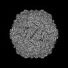 13164MC  9gotC  9hq7C 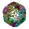 9hqcC M: map data used to model this data C: citing same article ( |
|---|---|
| Similar structure data |


-Links
 PDBj
PDBj
- Assembly
| Deposited unit | 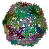
|
|---|---|
| 1 |
|
-Components
| #1: Protein | Mass: 28863.346 Da / Num. of mol.: 60 Source method: isolated from a genetically manipulated source Source: (gene. exp.) Mycobacterium tuberculosis (bacteria)Gene: cfp29, lin, C0094_04325, CAB90_00906, DSI38_23045, E5M05_20280, E5M52_19875, E5M78_19835, ERS007661_02029, ERS007665_01536, ERS007679_03499, ERS007688_03248, ERS007741_01530, ERS013471_03708, ...Gene: cfp29, lin, C0094_04325, CAB90_00906, DSI38_23045, E5M05_20280, E5M52_19875, E5M78_19835, ERS007661_02029, ERS007665_01536, ERS007679_03499, ERS007688_03248, ERS007741_01530, ERS013471_03708, ERS023446_03804, ERS027659_01280, ERS094182_03983, F6W99_03480, FRD82_02930, GCL30_20955, SAMEA2683035_02949 Production host: References: UniProt: A0A045HTX8, Hydrolases; Acting on peptide bonds (peptidases) Has protein modification | N | |
|---|
-Experimental details
-Experiment
| Experiment | Method: ELECTRON MICROSCOPY |
|---|---|
| EM experiment | Aggregation state: PARTICLE / 3D reconstruction method: single particle reconstruction |
- Sample preparation
| Component | Name: encapsulin shell from Mycobacterium tuberculosis / Type: ORGANELLE OR CELLULAR COMPONENT / Entity ID: all / Source: RECOMBINANT |
|---|---|
| Molecular weight | Value: 1.729 MDa / Experimental value: NO |
| Source (natural) | Organism: Mycobacterium tuberculosis H37Rv (bacteria) |
| Source (recombinant) | Organism: |
| Buffer solution | pH: 7 |
| Specimen | Conc.: 2.5 mg/ml / Embedding applied: NO / Shadowing applied: NO / Staining applied: NO / Vitrification applied: YES |
| Specimen support | Grid material: GOLD / Grid mesh size: 300 divisions/in. / Grid type: Quantifoil R1.2/1.3 |
| Vitrification | Instrument: FEI VITROBOT MARK IV / Cryogen name: ETHANE / Humidity: 90 % |
- Electron microscopy imaging
| Experimental equipment |  Model: Titan Krios / Image courtesy: FEI Company |
|---|---|
| Microscopy | Model: FEI TITAN KRIOS |
| Electron gun | Electron source:  FIELD EMISSION GUN / Accelerating voltage: 300 kV / Illumination mode: FLOOD BEAM FIELD EMISSION GUN / Accelerating voltage: 300 kV / Illumination mode: FLOOD BEAM |
| Electron lens | Mode: BRIGHT FIELD / Nominal magnification: 163000 X / Nominal defocus max: 2000 nm / Nominal defocus min: 700 nm |
| Specimen holder | Cryogen: NITROGEN / Specimen holder model: FEI TITAN KRIOS AUTOGRID HOLDER |
| Image recording | Average exposure time: 1.8 sec. / Electron dose: 40 e/Å2 / Film or detector model: GATAN K3 (6k x 4k) |
- Processing
| EM software |
| ||||||||||||||||||||||||||||||||
|---|---|---|---|---|---|---|---|---|---|---|---|---|---|---|---|---|---|---|---|---|---|---|---|---|---|---|---|---|---|---|---|---|---|
| CTF correction | Type: PHASE FLIPPING AND AMPLITUDE CORRECTION | ||||||||||||||||||||||||||||||||
| Particle selection | Num. of particles selected: 89072 | ||||||||||||||||||||||||||||||||
| Symmetry | Point symmetry: I (icosahedral) | ||||||||||||||||||||||||||||||||
| 3D reconstruction | Resolution: 2.29 Å / Resolution method: FSC 0.143 CUT-OFF / Num. of particles: 57805 / Symmetry type: POINT | ||||||||||||||||||||||||||||||||
| Atomic model building | B value: 34.36 / Protocol: AB INITIO MODEL / Space: REAL / Target criteria: multiple | ||||||||||||||||||||||||||||||||
| Atomic model building | PDB-ID: 619G Accession code: 619G / Source name: PDB / Type: experimental model |

