 Movie
Movie Controller

[English] 日本語
 Yorodumi
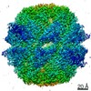
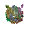
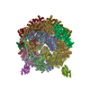
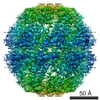
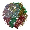
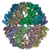
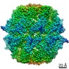
- PDB-7nvn: Human TRiC complex in closed state with nanobody and tubulin bound -
+ Open data

- Basic information
| Entry | Database: PDB / ID: 7nvn | ||||||
|---|---|---|---|---|---|---|---|
| Title | Human TRiC complex in closed state with nanobody and tubulin bound | ||||||
 Components Components |
| ||||||
 Keywords Keywords | CHAPERONE / TRiC / CCT / ATP hydrolysis / type II chaperonin / protein folding / tubulin | ||||||
| Function / homology |  Function and homology information Function and homology informationPost-chaperonin tubulin folding pathway / Cilium Assembly / Carboxyterminal post-translational modifications of tubulin / Microtubule-dependent trafficking of connexons from Golgi to the plasma membrane / positive regulation of protein localization to Cajal body / zona pellucida receptor complex / positive regulation of establishment of protein localization to telomere / scaRNA localization to Cajal body / positive regulation of telomerase RNA localization to Cajal body / chaperonin-containing T-complex ...Post-chaperonin tubulin folding pathway / Cilium Assembly / Carboxyterminal post-translational modifications of tubulin / Microtubule-dependent trafficking of connexons from Golgi to the plasma membrane / positive regulation of protein localization to Cajal body / zona pellucida receptor complex / positive regulation of establishment of protein localization to telomere / scaRNA localization to Cajal body / positive regulation of telomerase RNA localization to Cajal body / chaperonin-containing T-complex / BBSome-mediated cargo-targeting to cilium / tubulin complex assembly / Sealing of the nuclear envelope (NE) by ESCRT-III / Intraflagellar transport / Formation of tubulin folding intermediates by CCT/TriC / binding of sperm to zona pellucida / Folding of actin by CCT/TriC / Gap junction assembly / Kinesins / Prefoldin mediated transfer of substrate to CCT/TriC / Assembly and cell surface presentation of NMDA receptors / COPI-independent Golgi-to-ER retrograde traffic / RHOBTB1 GTPase cycle / COPI-dependent Golgi-to-ER retrograde traffic / WD40-repeat domain binding / pericentriolar material / Association of TriC/CCT with target proteins during biosynthesis / Recycling pathway of L1 / sperm head-tail coupling apparatus / chaperone-mediated protein complex assembly / RHOBTB2 GTPase cycle / RHO GTPases activate IQGAPs / Hydrolases; Acting on acid anhydrides; In phosphorus-containing anhydrides / beta-tubulin binding / Hedgehog 'off' state / intercellular bridge / COPI-mediated anterograde transport / Activation of AMPK downstream of NMDARs / positive regulation of telomere maintenance via telomerase / heterochromatin / MHC class II antigen presentation / protein folding chaperone / Recruitment of NuMA to mitotic centrosomes / Gene and protein expression by JAK-STAT signaling after Interleukin-12 stimulation / Mitotic Prometaphase / HSP90 chaperone cycle for steroid hormone receptors (SHR) in the presence of ligand / EML4 and NUDC in mitotic spindle formation / acrosomal vesicle / Resolution of Sister Chromatid Cohesion / Translocation of SLC2A4 (GLUT4) to the plasma membrane / mRNA 3'-UTR binding / ATP-dependent protein folding chaperone / RHO GTPases Activate Formins / cerebral cortex development / PKR-mediated signaling / structural constituent of cytoskeleton / microtubule cytoskeleton organization / mRNA 5'-UTR binding / neuron migration / response to virus / HCMV Early Events / Aggrephagy / azurophil granule lumen / mitotic spindle / The role of GTSE1 in G2/M progression after G2 checkpoint / : / melanosome / Separation of Sister Chromatids / mitotic cell cycle / Cooperation of PDCL (PhLP1) and TRiC/CCT in G-protein beta folding / G-protein beta-subunit binding / extracellular vesicle / protein folding / microtubule cytoskeleton / cell body / sperm midpiece / secretory granule lumen / ficolin-1-rich granule lumen / cytoskeleton / microtubule / protein stabilization / cadherin binding / GTPase activity / Neutrophil degranulation / ubiquitin protein ligase binding / centrosome / GTP binding / Golgi apparatus / ATP hydrolysis activity / RNA binding / extracellular exosome / extracellular region / nucleoplasm / ATP binding / metal ion binding / identical protein binding / nucleus / cytoplasm / cytosol Similarity search - Function | ||||||
| Biological species |  Homo sapiens (human) Homo sapiens (human) | ||||||
| Method | ELECTRON MICROSCOPY / single particle reconstruction / cryo EM / Resolution: 3 Å | ||||||
 Authors Authors | Kelly, J.J. / Chi, G. / Bulawa, C. / Paavilainen, V.O. / Bountra, C. / Huiskonen, J.T. / Yue, W. | ||||||
 Citation Citation | Journal: Nat Struct Mol Biol / Year: 2022 Title: Snapshots of actin and tubulin folding inside the TRiC chaperonin. Authors: John J Kelly / Dale Tranter / Els Pardon / Gamma Chi / Holger Kramer / Lotta Happonen / Kelly M Knee / Jay M Janz / Jan Steyaert / Christine Bulawa / Ville O Paavilainen / Juha T Huiskonen / Wyatt W Yue /      Abstract: The integrity of a cell's proteome depends on correct folding of polypeptides by chaperonins. The chaperonin TCP-1 ring complex (TRiC) acts as obligate folder for >10% of cytosolic proteins, ...The integrity of a cell's proteome depends on correct folding of polypeptides by chaperonins. The chaperonin TCP-1 ring complex (TRiC) acts as obligate folder for >10% of cytosolic proteins, including he cytoskeletal proteins actin and tubulin. Although its architecture and how it recognizes folding substrates are emerging from structural studies, the subsequent fate of substrates inside the TRiC chamber is not defined. We trapped endogenous human TRiC with substrates (actin, tubulin) and cochaperone (PhLP2A) at different folding stages, for structure determination by cryo-EM. The already-folded regions of client proteins are anchored at the chamber wall, positioning unstructured regions toward the central space to achieve their native fold. Substrates engage with different sections of the chamber during the folding cycle, coupled to TRiC open-and-close transitions. Further, the cochaperone PhLP2A modulates folding, acting as a molecular strut between substrate and TRiC chamber. Our structural snapshots piece together an emerging model of client protein folding within TRiC. | ||||||
| History |
|
- Structure visualization
| Movie |
Movie viewer |
|---|---|
| Structure viewer | Molecule: MolmilJmol/JSmol |
- Downloads & links
-Download
| PDBx/mmCIF format | 7nvn.cif.gz | 1.5 MB | Display | PDBx/mmCIF format |
|---|---|---|---|---|
| PDB format | pdb7nvn.ent.gz | 1.2 MB | Display | PDB format |
| PDBx/mmJSON format | 7nvn.json.gz | Tree view | PDBx/mmJSON format | |
| Others |  Other downloads Other downloads |
-Validation report
| Arichive directory | https://data.pdbj.org/pub/pdb/validation_reports/nv/7nvnftp://data.pdbj.org/pub/pdb/validation_reports/nv/7nvn | HTTPS FTP |
|---|
-Related structure data
| Related structure data | 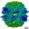 12607MC 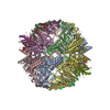 7nvlC 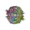 7nvmC  7nvoC C: citing same article ( M: map data used to model this data |
|---|---|
| Similar structure data |
-Links
 PDBj
PDBj


- Assembly
| Deposited unit | 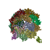
|
|---|---|
| 1 |
|
-Components
-T-complex protein 1 subunit ... , 8 types, 16 molecules AaBbDdEeGgHhQqZz
| #1: Protein | Mass: 60418.477 Da / Num. of mol.: 2 / Source method: isolated from a natural source / Source: (natural) Homo sapiens (human) / References: UniProt: P17987#2: Protein | Mass: 57567.141 Da / Num. of mol.: 2 / Source method: isolated from a natural source / Source: (natural) Homo sapiens (human) / References: UniProt: P78371#3: Protein | Mass: 57996.113 Da / Num. of mol.: 2 / Source method: isolated from a natural source / Source: (natural) Homo sapiens (human) / References: UniProt: P50991#4: Protein | Mass: 59749.957 Da / Num. of mol.: 2 Source method: isolated from a genetically manipulated source Source: (gene. exp.) Homo sapiens (human) / Gene: CCT5, CCTE, KIAA0098 / Production host: Homo sapiens (human) / References: UniProt: P48643#5: Protein | Mass: 60613.855 Da / Num. of mol.: 2 / Source method: isolated from a natural source / Source: (natural) Homo sapiens (human) / References: UniProt: P49368#6: Protein | Mass: 59443.535 Da / Num. of mol.: 2 / Source method: isolated from a natural source / Source: (natural) Homo sapiens (human) / References: UniProt: Q99832#8: Protein | Mass: 59691.422 Da / Num. of mol.: 2 / Source method: isolated from a natural source / Source: (natural) Homo sapiens (human) / References: UniProt: P50990#9: Protein | Mass: 58106.086 Da / Num. of mol.: 2 / Source method: isolated from a natural source / Source: (natural) Homo sapiens (human) / References: UniProt: P40227 |
|---|
-Antibody / Protein , 2 types, 3 molecules NnT
| #10: Protein | Mass: 49921.730 Da / Num. of mol.: 1 / Source method: isolated from a natural source / Source: (natural) Homo sapiens (human) / References: UniProt: Q13885 |
|---|---|
| #7: Antibody | Mass: 14412.816 Da / Num. of mol.: 2 Source method: isolated from a genetically manipulated source Source: (gene. exp.)  |
-Non-polymers , 4 types, 67 molecules 


| #11: Chemical | ChemComp-ADP /  Mass: 427.201 Da / Num. of mol.: 16 / Source method: obtained synthetically / Formula: C10H15N5O10P2 / Comment: ADP, energy-carrying molecule*YM Mass: 427.201 Da / Num. of mol.: 16 / Source method: obtained synthetically / Formula: C10H15N5O10P2 / Comment: ADP, energy-carrying molecule*YM#12: Chemical | ChemComp-MG /  Mass: 24.305 Da / Num. of mol.: 16 / Source method: obtained synthetically / Formula: Mg Mass: 24.305 Da / Num. of mol.: 16 / Source method: obtained synthetically / Formula: Mg#13: Chemical | ChemComp-AF3 /  Mass: 83.977 Da / Num. of mol.: 16 / Source method: obtained synthetically / Formula: AlF3 Mass: 83.977 Da / Num. of mol.: 16 / Source method: obtained synthetically / Formula: AlF3#14: Water | ChemComp-HOH / | Mass: 18.015 Da / Num. of mol.: 19 / Source method: isolated from a natural source / Formula: H2O |
|---|
-Details
| Has ligand of interest | N |
|---|---|
| Has protein modification | Y |
-Experimental details
-Experiment
| Experiment | Method: ELECTRON MICROSCOPY |
|---|---|
| EM experiment | Aggregation state: PARTICLE / 3D reconstruction method: single particle reconstruction |
- Sample preparation
| Component |
| ||||||||||||||||||||||||||||||
|---|---|---|---|---|---|---|---|---|---|---|---|---|---|---|---|---|---|---|---|---|---|---|---|---|---|---|---|---|---|---|---|
| Molecular weight |
| ||||||||||||||||||||||||||||||
| Source (natural) |
| ||||||||||||||||||||||||||||||
| Source (recombinant) |
| ||||||||||||||||||||||||||||||
| Buffer solution | pH: 7.5 | ||||||||||||||||||||||||||||||
| Specimen | Embedding applied: NO / Shadowing applied: NO / Staining applied: NO / Vitrification applied: YES | ||||||||||||||||||||||||||||||
| Vitrification | Cryogen name: ETHANE |
- Electron microscopy imaging
| Experimental equipment |  Model: Titan Krios / Image courtesy: FEI Company |
|---|---|
| Microscopy | Model: TFS KRIOS |
| Electron gun | Electron source:  FIELD EMISSION GUN / Accelerating voltage: 300 kV / Illumination mode: FLOOD BEAM FIELD EMISSION GUN / Accelerating voltage: 300 kV / Illumination mode: FLOOD BEAM |
| Electron lens | Mode: BRIGHT FIELD |
| Image recording | Electron dose: 43 e/Å2 / Film or detector model: GATAN K2 SUMMIT (4k x 4k) |
- Processing
| Software |
| ||||||||||||||||||||||||
|---|---|---|---|---|---|---|---|---|---|---|---|---|---|---|---|---|---|---|---|---|---|---|---|---|---|
| CTF correction | Type: PHASE FLIPPING AND AMPLITUDE CORRECTION | ||||||||||||||||||||||||
| Symmetry | Point symmetry: C1 (asymmetric) | ||||||||||||||||||||||||
| 3D reconstruction | Resolution: 3 Å / Resolution method: FSC 0.143 CUT-OFF / Num. of particles: 93758 / Symmetry type: POINT | ||||||||||||||||||||||||
| Refinement | Cross valid method: NONE Stereochemistry target values: GeoStd + Monomer Library + CDL v1.2 | ||||||||||||||||||||||||
| Displacement parameters | Biso mean: 73.26 Å2 | ||||||||||||||||||||||||
| Refine LS restraints |
|
