 Movie
Movie Controller


+ Open data

- Basic information
| Entry | Database: PDB / ID: 7nd2 | ||||||
|---|---|---|---|---|---|---|---|
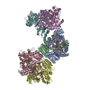
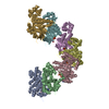
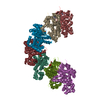
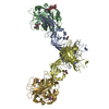
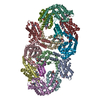
| Title | Cryo-EM structure of the human FERRY complex | ||||||
 Components Components |
| ||||||
 Keywords Keywords | RNA BINDING PROTEIN / Human FERRY complex / Five-subunit Early endosome RNA and Ribosome intermediarY complex / Intracellular RNA transport / Early Endosome-associated transport of RNA | ||||||
| Function / homology |  Function and homology information Function and homology informationquinone metabolic process / glyoxalase III activity / quinone reductase (NADPH) activity / methylglyoxal catabolic process to D-lactate via S-lactoyl-glutathione / Oxidoreductases / NADP binding / early endosome / RNA binding / extracellular exosome / identical protein binding ...quinone metabolic process / glyoxalase III activity / quinone reductase (NADPH) activity / methylglyoxal catabolic process to D-lactate via S-lactoyl-glutathione / Oxidoreductases / NADP binding / early endosome / RNA binding / extracellular exosome / identical protein binding / membrane / cytoplasm / cytosol Similarity search - Function | ||||||
| Biological species |  Homo sapiens (human) Homo sapiens (human) | ||||||
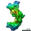
| Method | ELECTRON MICROSCOPY / single particle reconstruction / cryo EM / Resolution: 4 Å | ||||||
 Authors Authors | Quentin, D. / Klink, B.U. / Raunser, S. | ||||||
| Funding support | 1items
| ||||||
 Citation Citation | Journal: Mol Cell / Year: 2023 Title: Structural basis of mRNA binding by the human FERRY Rab5 effector complex. Authors: Dennis Quentin / Jan S Schuhmacher / Björn U Klink / Jeni Lauer / Tanvir R Shaikh / Pim J Huis In 't Veld / Luisa M Welp / Henning Urlaub / Marino Zerial / Stefan Raunser /  Abstract: The pentameric FERRY Rab5 effector complex is a molecular link between mRNA and early endosomes in mRNA intracellular distribution. Here, we determine the cryo-EM structure of human FERRY. It reveals ...The pentameric FERRY Rab5 effector complex is a molecular link between mRNA and early endosomes in mRNA intracellular distribution. Here, we determine the cryo-EM structure of human FERRY. It reveals a unique clamp-like architecture that bears no resemblance to any known structure of Rab effectors. A combination of functional and mutational studies reveals that while the Fy-2 C-terminal coiled-coil acts as binding region for Fy-1/3 and Rab5, both coiled-coils and Fy-5 concur to bind mRNA. Mutations causing truncations of Fy-2 in patients with neurological disorders impair Rab5 binding or FERRY complex assembly. Thus, Fy-2 serves as a binding hub connecting all five complex subunits and mediating the binding to mRNA and early endosomes via Rab5. Our study provides mechanistic insights into long-distance mRNA transport and demonstrates that the particular architecture of FERRY is closely linked to a previously undescribed mode of RNA binding, involving coiled-coil domains. | ||||||
| History |
|
- Structure visualization
| Movie |
Movie viewer |
|---|---|
| Structure viewer | Molecule: MolmilJmol/JSmol |
- Downloads & links
-Download
| PDBx/mmCIF format | 7nd2.cif.gz | 395.5 KB | Display | PDBx/mmCIF format |
|---|---|---|---|---|
| PDB format | pdb7nd2.ent.gz | 311.2 KB | Display | PDB format |
| PDBx/mmJSON format | 7nd2.json.gz | Tree view | PDBx/mmJSON format | |
| Others |  Other downloads Other downloads |
-Validation report
| Summary document | 7nd2_validation.pdf.gz | 789.3 KB | Display | wwPDB validaton report |
|---|---|---|---|---|
| Full document | 7nd2_full_validation.pdf.gz | 825.9 KB | Display | |
| Data in XML | 7nd2_validation.xml.gz | 62.6 KB | Display | |
| Data in CIF | 7nd2_validation.cif.gz | 95.8 KB | Display | |
| Arichive directory | https://data.pdbj.org/pub/pdb/validation_reports/nd/7nd2ftp://data.pdbj.org/pub/pdb/validation_reports/nd/7nd2 | HTTPS FTP |
-Related structure data
| Related structure data |  12273MC 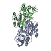 8a3oC 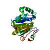 8a3pC M: map data used to model this data C: citing same article ( |
|---|---|
| Similar structure data |
-Links
 PDBj
PDBj


- Assembly
| Deposited unit | 
|
|---|---|
| 1 |
|
-Components
| #1: Protein | Mass: 88782.836 Da / Num. of mol.: 2 Source method: isolated from a genetically manipulated source Source: (gene. exp.) Homo sapiens (human) / Gene: PPP1R21, CCDC128, KLRAQ1 / Production host:   Spodoptera frugiperda (fall armyworm) / Strain (production host): Sf-9 / References: UniProt: Q6ZMI0 Spodoptera frugiperda (fall armyworm) / Strain (production host): Sf-9 / References: UniProt: Q6ZMI0#2: Protein | Mass: 39661.414 Da / Num. of mol.: 2 Source method: isolated from a genetically manipulated source Details: N-terminal His-6 tag / Source: (gene. exp.) Homo sapiens (human) / Gene: CRYZL1, 4P11 / Production host:  #3: Protein | Mass: 24237.488 Da / Num. of mol.: 4 Source method: isolated from a genetically manipulated source Source: (gene. exp.) Homo sapiens (human) / Gene: GATD1, PDDC1 / Production host: |
|---|
-Experimental details
-Experiment
| Experiment | Method: ELECTRON MICROSCOPY |
|---|---|
| EM experiment | Aggregation state: PARTICLE / 3D reconstruction method: single particle reconstruction |
- Sample preparation
| Component |
| ||||||||||||||||||||||||||||||
|---|---|---|---|---|---|---|---|---|---|---|---|---|---|---|---|---|---|---|---|---|---|---|---|---|---|---|---|---|---|---|---|
| Source (natural) |
| ||||||||||||||||||||||||||||||
| Source (recombinant) |
| ||||||||||||||||||||||||||||||
| Buffer solution | pH: 7.5 | ||||||||||||||||||||||||||||||
| Buffer component |
| ||||||||||||||||||||||||||||||
| Specimen | Conc.: 0.7 mg/ml / Embedding applied: NO / Shadowing applied: NO / Staining applied: NO / Vitrification applied: YES | ||||||||||||||||||||||||||||||
| Specimen support | Grid material: GOLD / Grid mesh size: 300 divisions/in. / Grid type: UltrAuFoil R1.2/1.3 | ||||||||||||||||||||||||||||||
| Vitrification | Instrument: FEI VITROBOT MARK III / Cryogen name: ETHANE / Humidity: 100 % / Chamber temperature: 286 K / Details: 3s blotting time |
- Electron microscopy imaging
| Experimental equipment |  Model: Titan Krios / Image courtesy: FEI Company |
|---|---|
| Microscopy | Model: TFS KRIOS |
| Electron gun | Electron source:  FIELD EMISSION GUN / Accelerating voltage: 300 kV / Illumination mode: FLOOD BEAM FIELD EMISSION GUN / Accelerating voltage: 300 kV / Illumination mode: FLOOD BEAM |
| Electron lens | Mode: BRIGHT FIELD / Calibrated defocus min: 1600 nm / Calibrated defocus max: 2800 nm |
| Specimen holder | Cryogen: NITROGEN / Specimen holder model: FEI TITAN KRIOS AUTOGRID HOLDER |
| Image recording | Average exposure time: 15 sec. / Electron dose: 75.8 e/Å2 / Film or detector model: GATAN K2 SUMMIT (4k x 4k) / Num. of real images: 1879 |
| EM imaging optics | Energyfilter name: GIF Bioquantum / Energyfilter slit width: 20 eV |
| Image scans | Movie frames/image: 40 |
- Processing
| Software | Name: PHENIX / Version: 1.18.2_3874: / Classification: refinement | |||||||||||||||||||||||||||||||||
|---|---|---|---|---|---|---|---|---|---|---|---|---|---|---|---|---|---|---|---|---|---|---|---|---|---|---|---|---|---|---|---|---|---|---|
| EM software |
| |||||||||||||||||||||||||||||||||
| CTF correction | Type: PHASE FLIPPING AND AMPLITUDE CORRECTION | |||||||||||||||||||||||||||||||||
| Particle selection | Num. of particles selected: 1800000 | |||||||||||||||||||||||||||||||||
| Symmetry | Point symmetry: C2 (2 fold cyclic) | |||||||||||||||||||||||||||||||||
| 3D reconstruction | Resolution: 4 Å / Resolution method: FSC 0.143 CUT-OFF / Num. of particles: 18300 / Symmetry type: POINT | |||||||||||||||||||||||||||||||||
| Atomic model building | Details: To build the model for the (CRYZL1)2(PPP1r21)2(GATD1)4 core of the FERRY complex, the obtained crystal structures of CRYZL1 and GATD1 were initially fitted into the corresponding density ...Details: To build the model for the (CRYZL1)2(PPP1r21)2(GATD1)4 core of the FERRY complex, the obtained crystal structures of CRYZL1 and GATD1 were initially fitted into the corresponding density using the rigid body fitting tool in Chimera. trRosetta, a de novo protein structure prediction algorithm that is based on direct energy minimization with restrained Rosetta, was used to obtain initial models for PPP1r21. The predicted model for the 6-helix bundle domain, containing residues 246 to 498, that matched our experimental density best was subsequently fitted similar as CRYZL1 and GATD1 using rigid body fit. Manual model building for the regions N- and C-terminal 6-helix bundle, which comprise residues 218 to 245 and 499 to 552, respectively, was further guided by secondary structure predictions of individual trRosetta runs for these regions, that include the vertical helix as well as the beginning of the two terminal coiled-coils of PPP1r21. With the resulting combined model, containing residues 2 to 349, 218 to 552 and 8 to 217 of CRYZL1, PPP1r21 and GATD1, respectively, a restrained refinement in PHENIX was performed. In the next step, the model was further refined using a combination of manual building in COOT and real-space refinement in PHENIX. | |||||||||||||||||||||||||||||||||
| Refine LS restraints |
|
